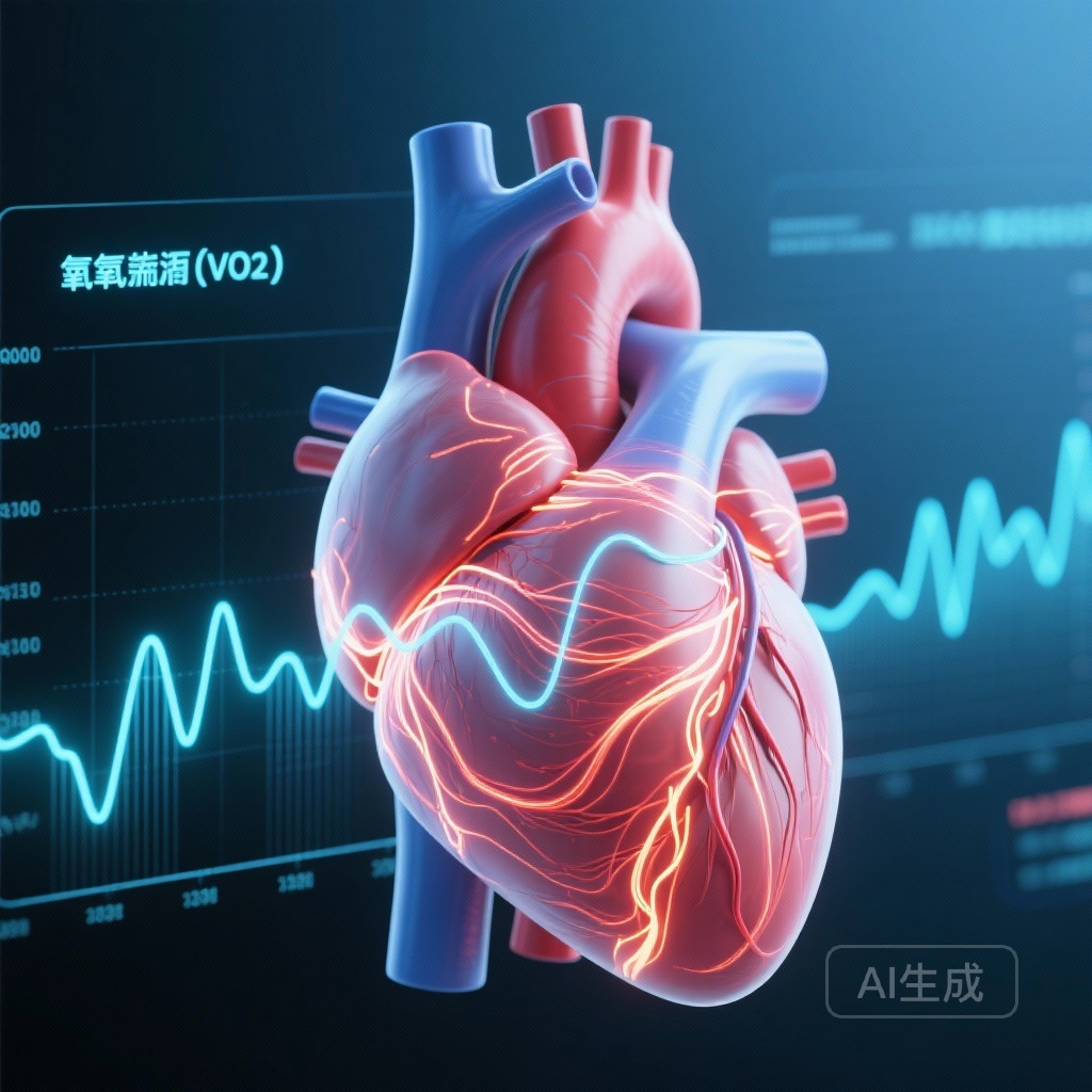
Introduction: Beyond Peak VO2 in Hypertrophic Cardiomyopathy
For decades, peak oxygen uptake (peak VO2) has served as the gold standard for assessing functional capacity and prognosis in patients with heart failure and hypertrophic cardiomyopathy (HCM). However, peak VO2 is highly dependent on patient effort and peripheral factors, which can sometimes obscure the primary contribution of cardiac performance. As clinicians seek more granular insights into myocardial efficiency, the recovery phase of exercise—specifically the kinetics of oxygen uptake recovery (VO2Rec)—has emerged as a promising frontier.
In patients with obstructive hypertrophic cardiomyopathy (oHCM), the pathophysiological hallmark is left ventricular outflow tract (LVOT) obstruction, leading to elevated filling pressures and impaired cardiac output reserve. While treatment with cardiac myosin inhibitors like aficamten has been shown to improve peak capacity, understanding how these therapies alter the heart’s ability to recover from exertion provides a more comprehensive view of hemodynamic restoration. This substudy of the SEQUOIA-HCM trial, published in Circulation, introduces VO2T12.5% as a simple yet powerful metric for this purpose.
High-Level Highlights
- VO2T12.5% is established as a novel, easily derived measure of oxygen recovery kinetics that correlates strongly with invasive hemodynamic markers of cardiac performance.
- Longer VO2T12.5% times (≥35 seconds) are independently associated with a significantly higher risk of heart failure hospitalization and death.
- Treatment with the cardiac myosin inhibitor aficamten significantly shortens VO2T12.5%, reflecting a direct improvement in cardiac function and symptomatic recovery.
- The metric offers a standardized way to evaluate exercise physiology that is less dependent on peak effort than traditional peak VO2 measurements.
The Physiological Basis of VO2 Recovery Kinetics
VO2 recovery reflects the rate at which the body clears the oxygen debt incurred during exercise. In a healthy heart, the cessation of exercise is followed by a rapid decline in VO2 as cardiac output and pulmonary pressures normalize. In advanced heart failure or symptomatic oHCM, this process is delayed. The delay is primarily driven by the inability of the heart to rapidly reduce filling pressures and maintain adequate stroke volume in the immediate post-exercise period.
The researchers focused on VO2T12.5%—the time required for VO2 to decline by 12.5% from its peak value. Unlike total recovery time, which can be influenced by metabolic clearance of lactic acid, this early phase of recovery is more closely tethered to central cardiac hemodynamics and the immediate restoration of efficient circulation.
Study Design: The MGH-ExS and SEQUOIA-HCM Cohorts
The study was conducted in two distinct phases. First, to establish the physiological and prognostic validity of VO2Rec, the investigators analyzed the Massachusetts General Hospital Exercise Study (MGH-ExS) cohort. This included 814 patients referred for dyspnea on exertion who underwent cardiopulmonary exercise testing (CPET) combined with invasive hemodynamic monitoring. This allowed the team to correlate VO2 kinetics with measured pulmonary capillary wedge pressure (PCWP) and cardiac output (CO).
The second phase applied these findings to the SEQUOIA-HCM trial (Safety, Efficacy, and Quantitative Understanding of Obstruction Impact of Aficamten in Hypertrophic Cardiomyopathy). This was a pivotal phase 3, randomized, double-blind, placebo-controlled trial involving 263 participants with symptomatic oHCM. Patients were randomized to receive either aficamten or placebo for 24 weeks. The primary focus of this substudy was to determine if aficamten-mediated reductions in LVOT obstruction translated into improved (shortened) VO2T12.5% times.
Key Findings: VO2T12.5% as a Window into Cardiac Performance
Hemodynamic Correlations and Prognostic Utility
Data from the MGH-ExS cohort provided robust evidence that VO2T12.5% is a cardiospecific marker. Patients with a VO2T12.5% of 35 seconds or longer demonstrated a significantly steeper PCWP-to-CO slope (P < 0.0001), indicating worse hemodynamic efficiency during exercise. Interestingly, there was no significant difference in peripheral oxygen extraction between those with fast and slow recovery, reinforcing that the delay is a cardiac, rather than a peripheral muscular, issue.
Furthermore, the prognostic value was striking. For every 15-second increase in VO2T12.5%, the hazard ratio for the composite endpoint of heart failure hospitalization or all-cause mortality was 1.54 (95% CI, 1.35–1.76; P < 0.001). This suggests that how a patient recovers from exercise may be just as important as how much work they can perform at peak.
The Impact of Aficamten on Recovery Kinetics
In the SEQUOIA-HCM cohort, the baseline VO2T12.5% was 45 ± 20 seconds. After 24 weeks of treatment, patients receiving aficamten saw their recovery time shorten by an average of 8 seconds compared to the placebo group (95% CI, -12 to -5 seconds; P < 0.001).
The clinical significance was highlighted by the Odds Ratio (OR): patients on aficamten were 3.7 times more likely to achieve a clinically meaningful improvement (defined as a shortening of ≥15 seconds) in VO2T12.5% compared to those on placebo. The number needed to treat (NNT) to achieve this improvement was remarkably low at 4.8. These improvements in recovery kinetics correlated precisely with reductions in NT-proBNP, cardiac troponin I, and the LVOT gradient, suggesting that the drug’s mechanism of reducing myocardial wall stress and obstruction directly facilitates faster physiological recovery.
Expert Commentary: Mechanistic Insights and Clinical Integration
The introduction of VO2T12.5% addresses a critical gap in CPET interpretation. While peak VO2 remains vital, it is often limited by a patient’s orthopedic constraints, motivation, or general deconditioning. Recovery kinetics, particularly in the first 30 to 60 seconds post-exercise, are less susceptible to these confounders.
The fact that VO2T12.5% improved in tandem with biomarkers like NT-proBNP and troponin provides strong biological plausibility. By inhibiting the excessive actin-myosin cross-bridging characteristic of HCM, aficamten improves diastolic relaxation and reduces the obstructive gradient. This study confirms that these cellular and anatomical changes result in a measurable improvement in how the entire circulatory system resets after stress. Clinicians should consider incorporating VO2 recovery metrics into routine CPET reports to better phenotype their patients’ cardiac reserve.
Conclusion: A New Standard for Exercise Assessment?
The SEQUOIA-HCM substudy successfully validates VO2T12.5% as a novel, reproducible, and clinically meaningful metric in the management of oHCM. It bridges the gap between invasive hemodynamics and non-invasive exercise testing, providing a clear signal of cardiac performance. As aficamten and other cardiac myosin inhibitors reshape the treatment landscape for HCM, VO2T12.5% offers a sensitive tool for monitoring therapeutic response and predicting long-term outcomes. Its simplicity ensures that it can be easily integrated into existing CPET protocols without the need for additional equipment or complex calculations.
Funding and Clinical Trial Information
The SEQUOIA-HCM trial was funded by Cytokinetics, Inc. ClinicalTrials.gov Identifier: NCT05186818.
References
1. Campain J, et al. Characterization and Application of Novel Exercise Recovery Patterns That Reflect Cardiac Performance: A Substudy of the SEQUOIA-HCM Trial. Circulation. 2025;152(14):990-1002. doi:10.1161/CIRCULATIONAHA.124.073585.
2. Maron MS, et al. Aficamten for Symptomatic Obstructive Hypertrophic Cardiomyopathy: Results of the SEQUOIA-HCM Phase 3 Trial. J Am Coll Cardiol. 2024.
3. Malhotra R, et al. Cardiopulmonary Exercise Testing in Heart Failure. JACC: Heart Failure. 2016;4(8):607-616.