Introduction: The Challenge of Early Prognostication in Neonatal HIE
Hypoxic-ischemic encephalopathy (HIE) remains a leading cause of neonatal mortality and long-term neurodevelopmental disability worldwide. While the introduction of therapeutic hypothermia (TH) has revolutionized management, reducing the risk of death or severe disability, a significant proportion of treated infants still experience adverse outcomes. For clinicians and families, the ability to accurately predict long-term neurodevelopmental trajectories within the first 24 to 48 hours of life is critical. Early and precise risk stratification not only informs clinical decision-making regarding the intensity of care but also serves as a vital tool for selecting participants in clinical trials for adjunctive neuroprotective therapies.
Traditional bedside assessments, such as the Sarnat score, provide immediate clinical insight but are often limited by inter-rater variability and the confounding effects of sedation or therapeutic hypothermia. While electroencephalography (EEG) is recognized as the gold standard for monitoring brain function in the NICU, its utility is frequently hampered by the need for specialized pediatric neurophysiologists to interpret complex background patterns in real-time. The emergence of automated EEG analysis offers a potential solution, providing objective, continuous, and rapid assessment of brain states.
The Role of EEG Background in Neonatal Encephalopathy
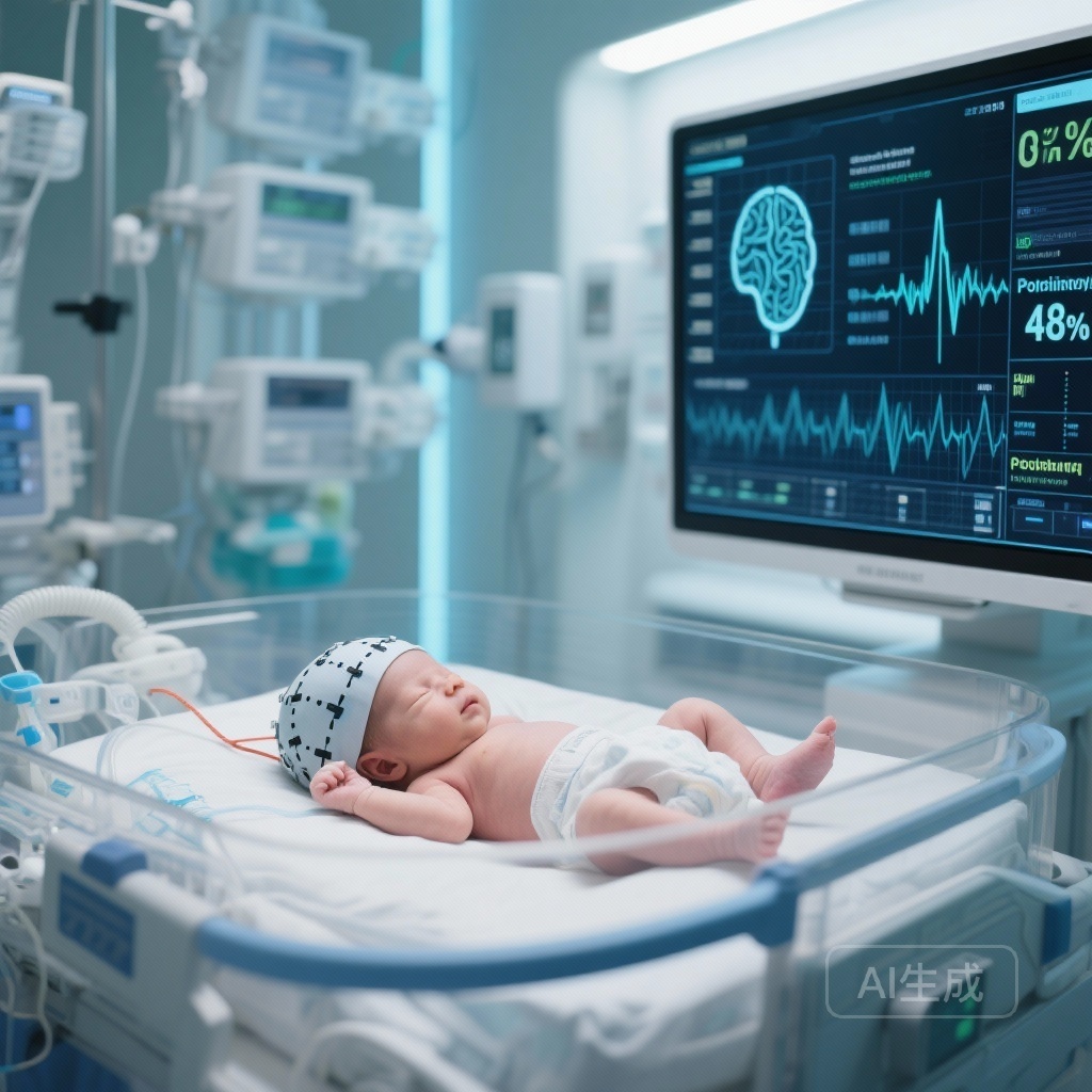
EEG background patterns reflect the functional integrity of the neonatal brain. In the context of HIE, the severity of background abnormality—ranging from mild discontinuity to burst suppression or a flat trace—correlates strongly with the extent of brain injury. However, interpreting these patterns requires significant expertise. The ‘Brain State of the Newborn’ (BSN) score was developed as a quantitative, automated tool to classify these background patterns into a simplified numerical scale. By leveraging signal processing and machine learning, the BSN score aims to provide a standardized metric that can be easily integrated into clinical practice, even in centers without 24-hour access to expert neurophysiologists.
Study Design and Methodology: A Secondary Analysis of the HEAL Trial
The study conducted by Cornet et al., published in JAMA Network Open, represents a significant step forward in validating automated EEG tools. This cohort study was a secondary analysis of the High-Dose Erythropoietin for Asphyxia and Encephalopathy (HEAL) trial, a multi-center randomized controlled trial that evaluated the efficacy of erythropoietin as an adjunctive therapy to therapeutic hypothermia.
The analysis included 203 neonates born at 36 weeks’ gestation or later who met the criteria for HIE and underwent therapeutic hypothermia. Raw EEG data were collected from nine academic centers across the United States, starting within the first 24 hours of life. The BSN scores were computed via a cloud-based service, providing a median score for each infant based on their EEG background. The primary outcome was a composite of death or severe neurodevelopmental impairment (NDI) at age 2 years, defined by standardized assessments including the Bayley Scales of Infant and Toddler Development, Third Edition (Bayley-III).
Key Findings: Enhancing Prognostic Accuracy
The results of the study underscore the potent estimative ability of automated EEG analysis. Among the 203 infants analyzed, 21 (10.3%) experienced severe NDI and 28 (13.8%) died by the age of 2 years.
Correlation with Expert Interpretation
One of the primary objectives was to determine if the BSN score accurately reflected the assessment of expert human readers. The researchers found a strong Pearson correlation coefficient of 0.69 (95% CI, 0.64-0.73) between the median BSN scores and expert-classified EEG background categories. This suggests that the automated algorithm is highly consistent with the ‘ground truth’ provided by experienced neurologists.
Superiority Over Clinical Variables
When evaluating the ability to predict the composite outcome of death or severe NDI, clinical variables alone (such as birth weight, pH, and Apgar scores) yielded an area under the receiver operating characteristic curve (AUROC) of 0.79 (95% CI, 0.70-0.87). However, the addition of the median overall BSN score significantly improved the model’s performance, raising the AUROC to 0.90 (95% CI, 0.84-0.97).
Furthermore, when the median BSN score at all available time points was included in the model, the AUROC reached a remarkable 0.93 (95% CI, 0.88-0.98). This indicates that the BSN score provides substantial incremental value beyond traditional clinical markers, offering a much higher degree of confidence in early-life prognosis.
Comparison to Expert Readers
Importantly, the prognostic accuracy of the automated BSN score was found to be statistically similar to the prognostic accuracy of expert assessments of the EEG background (AUROC, 0.90; 95% CI, 0.81-0.98). This finding is particularly relevant for clinical settings where expert neurology consultation is not immediately available, suggesting that automated tools could effectively fill the gap in expertise.
Clinical Implications and Translational Insight
The ability of the BSN score to match expert performance suggests it could be a transformative tool in the NICU. For many community hospitals or even regional centers, providing 24/7 expert EEG interpretation is logistically and financially challenging. An automated, objective score allows for real-time monitoring of brain recovery or deterioration, which can guide clinicians in communicating with families and planning long-term follow-up.
From a research perspective, the BSN score offers a standardized method for risk stratification. In future clinical trials for HIE, investigators could use the BSN score to identify infants at the highest risk for poor outcomes, ensuring that neuroprotective interventions are tested in the populations most likely to benefit. This reduces the ‘noise’ in clinical data and increases the power of the study to detect meaningful treatment effects.
Expert Commentary: Strengths and Limitations
While the findings are promising, several considerations must be addressed. The study is a secondary analysis of the HEAL trial, which, although robust, may be subject to selection bias as only infants with available raw EEG data were included. Additionally, while the BSN score correlates well with expert readers, it is not a replacement for comprehensive clinical assessment. The EEG background is just one piece of the puzzle; other factors, such as neuroimaging (MRI) and serial clinical examinations, remain essential components of the prognostic workup.
One potential limitation of the BSN score is its dependence on signal quality. In a busy NICU environment, electrical interference and movement artifacts can degrade EEG signals. Future iterations of automated tools will need to demonstrate continued reliability in the face of such common technical challenges. Moreover, the study focused on 2-year outcomes; longer-term follow-up into school age is necessary to determine if the BSN score can predict more subtle cognitive or behavioral deficits that may not be apparent in early childhood.
Conclusion: A New Era of Objective Neonatal Neurology
The study by Cornet and colleagues provides compelling evidence that automated EEG background analysis is a feasible and highly accurate method for predicting 2-year neurodevelopmental outcomes in neonates with HIE. By providing an objective score that rivals expert interpretation, the BSN tool has the potential to democratize high-level neurological monitoring. As we move toward more personalized and data-driven neonatal care, such automated systems will likely become indispensable in our efforts to minimize the burden of brain injury in our most vulnerable patients.
Funding and ClinicalTrials.gov
This study was supported by grants from the National Institute of Neurological Disorders and Stroke (NINDS). The HEAL trial is registered at ClinicalTrials.gov (NCT02816632). Additional support was provided by the University of California, San Francisco, and participating academic institutions.
References
1. Cornet MC, Numis AL, Wusthoff CJ, et al. Automated EEG Background Analysis and 2-Year Outcomes in Neonatal Hypoxic-Ischemic Encephalopathy. JAMA Netw Open. 2025;8(12):e2548321. doi:10.1001/jamanetworkopen.2025.48321.
2. Wu YW, Mayock DE, Kroon K, et al. High-Dose Erythropoietin for Asphyxia and Encephalopathy (HEAL): A Randomized Trial. N Engl J Med. 2022;387(2):148-159.
3. Tich SN, d’Allest AM. Predictive value of EEG in neonatal encephalopathy. Neurophysiol Clin. 2016;46(4-5):249-265.
4. Glass HC, Wusthoff CJ, Shellhaas RA. Amplitude-integrated EEG in the neonatal intensive care unit: use, interpretation, and pitfalls. J Perinatol. 2013;33(11):831-839.