Executive Highlights
Sarcoidosis, a multi-systemic granulomatous disorder, frequently manifests in the eye, presenting a significant challenge for clinicians managing inflammatory recurrence and long-term visual health. A large-scale multicentric study has provided new insights into the clinical trajectory of sarcoid uveitis. The key takeaways include:
- Approximately 66.7% of patients with sarcoid uveitis experience at least one ocular relapse during follow-up.
- The median time to the first ocular relapse is 2.6 years, emphasizing the need for long-term vigilance.
- Independent risk factors for shorter relapse-free survival include the presence of macular edema and persistent inflammation at the time of diagnosis.
- The visual prognosis is generally positive; while 6.5% of patients experienced some level of visual impairment, only 0.9% reached the threshold of legal blindness after a median follow-up of 7.8 years.
- Glaucoma and macular edema remain the primary drivers of permanent visual morbidity in this population.
The Burden of Sarcoid Uveitis in Clinical Practice
Sarcoidosis is characterized by the formation of non-caseating granulomas in various organs, with ocular involvement occurring in 25% to 50% of patients depending on the cohort and ethnicity. Uveitis is the most common ocular manifestation and can lead to severe complications if not managed aggressively. For the clinician, the primary goals are twofold: maintaining quiescent disease to prevent structural damage and preserving visual acuity.
Despite advances in immunosuppressive therapy and the introduction of biologic agents, many patients suffer from a relapsing-remitting course. Understanding which patients are at the highest risk for relapse and visual decline is essential for personalizing treatment intensity and monitoring frequency. The study by Abramowicz et al., published in Ophthalmology, addresses these clinical gaps by analyzing a large, well-characterized cohort over an extended period.
Study Design and Patient Demographics
This research utilized a multicentric, retrospective cohort design involving 336 patients. The demographic profile reflected a typical sarcoidosis population, with a female predominance (61.9%) and a median age at diagnosis of 52 years. Notably, the study achieved a high rate of diagnostic certainty, with sarcoidosis histologically proven in 83.0% of the participants.
The primary endpoints were the time to ocular relapse and the time to the development of visual impairment. The researchers utilized Kaplan-Meier curves to estimate relapse-free survival (RFS) and Cox proportional hazards models to identify specific variables associated with these outcomes. The median follow-up duration was 7.8 years, providing a robust window into the long-term natural history and treatment response of the disease.
Key Findings: Relapse Patterns and Risk Factors
The study found that ocular relapses are the rule rather than the exception in sarcoid uveitis. With two-thirds of the cohort experiencing a relapse, the data suggests that sarcoidosis in the eye is often a chronic, recurring condition rather than a monophasic event. The median time to relapse was relatively short at 2.6 years, but the interquartile range (1.6-4.4 years) indicates significant variability among patients.
The Impact of Baseline Macular Edema
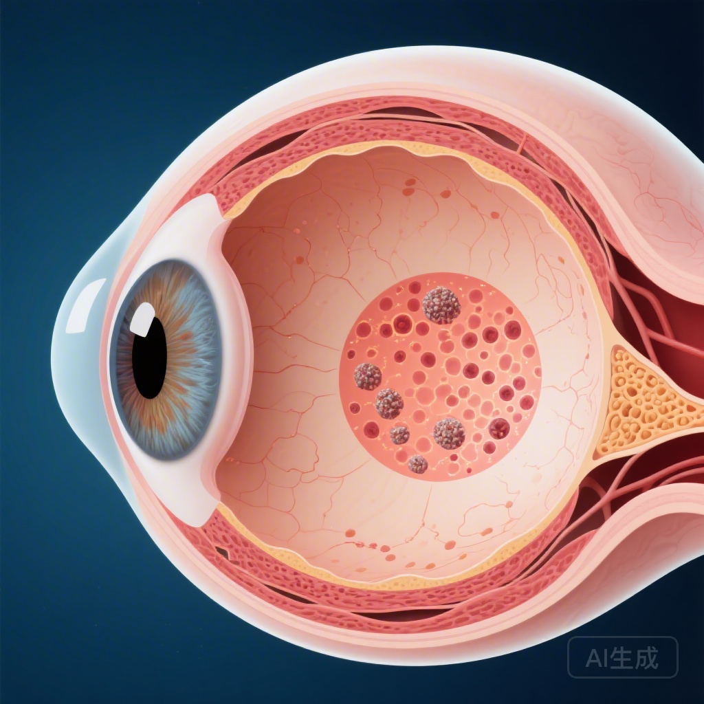
One of the most significant findings was the association between baseline macular edema and a higher hazard of relapse (HR 1.47). Macular edema is a common complication of posterior and panuveitis, representing a breakdown of the blood-retinal barrier. Its presence at diagnosis likely reflects a more aggressive inflammatory phenotype or a specific cytokine profile that predisposes the eye to recurrent bouts of inflammation.
Persistent Inflammation as a Red Flag
Persistent inflammation at baseline was even more strongly associated with relapse risk (HR 1.68). In this context, persistent inflammation refers to the inability to achieve complete quiescence within the initial treatment phase. This finding underscores the importance of achieving early and total control of intraocular inflammation. Clinicians may need to consider earlier escalation to second-line immunosuppressants or biologics in patients who do not respond rapidly to corticosteroids.
Visual Prognosis: Reassurance and Risk
Perhaps the most encouraging aspect of the study is the long-term visual outcome. Despite the high rate of relapse, 93.5% of patients maintained vision that did not meet the World Health Organization (WHO) criteria for visual impairment at the end of the follow-up period. Only 0.9% of patients became blind.
Causes of Vision Loss
When visual impairment did occur, it was primarily due to structural complications rather than active inflammation itself. The main causes identified were:
- Glaucoma (45.0%): Often a result of both the disease process (trabeculitis or synechiae) and the side effects of prolonged corticosteroid use.
- Macular Edema (40.0%): Highlighting its role as both a predictor of relapse and a direct cause of vision loss.
- Epiretinal Membrane (30.0%): A common secondary complication of chronic intraocular inflammation.
The fact that glaucoma was the leading cause of impairment serves as a critical reminder for ophthalmologists to monitor intraocular pressure vigilantly and employ steroid-sparing strategies whenever possible.
Expert Commentary and Clinical Implications
The findings from this multicentric study reinforce the necessity of a proactive and personalized approach to sarcoid uveitis. The association between early macular edema and future relapses suggests that these patients might benefit from more intensive initial therapy. Furthermore, the link between persistent inflammation and relapse highlights the inadequacy of “tolerating” low-grade inflammation; the goal should always be complete suppression of the inflammatory response.
However, the study’s retrospective nature does present limitations. Treatment protocols were not standardized across the different centers, which could influence the relapse rates. Additionally, the small number of visual impairment events, while positive for patients, made it difficult to statistically model the specific risk factors for vision loss. Future prospective studies utilizing standardized treatment algorithms and modern imaging, such as optical coherence tomography angiography (OCTA), may further refine our ability to predict outcomes.
From a multidisciplinary perspective, the high rate of histological confirmation in this study (83%) is commendable. It underscores the importance of collaboration between ophthalmologists, pulmonologists, and internal medicine specialists to confirm the systemic diagnosis, which in turn informs the long-term management strategy.
Conclusion
In conclusion, while sarcoid uveitis is characterized by a high frequency of ocular relapse, the long-term visual prognosis is excellent for the vast majority of patients. By identifying macular edema and persistent inflammation as key risk factors for recurrence, this study provides clinicians with valuable tools for risk stratification. Management should focus on achieving rapid quiescence, managing secondary complications like glaucoma, and maintaining long-term follow-up to catch relapses early, thereby ensuring that the “usable vision” of patients is preserved for decades.
References
1. Abramowicz S, Jacquot R, Siriphanh A, et al. Factors Associated with Ocular Relapse and Visual Prognosis in Sarcoid Uveitis. Ophthalmology. 2025;133(3):378-386. doi:10.1016/j.ophtha.2024.09.025 (Refers to PMID: 41177360).
2. Jabs DA, Nussenblatt RB, Rosenbaum JT. Standardization of uveitis nomenclature for reporting clinical data. Results of the First International Workshop. Am J Ophthalmol. 2005;140(3):509-516.
3. Jamilloux Y, Kodjikian L, Broussolle C, Sève P. Sarcoidosis and uveitis. Autoimmun Rev. 2014;13(12):1220-1227.