Highlight
- Right anomalous aortic origin of a coronary artery (R-AAOCA) is increasingly diagnosed but remains a rare congenital anomaly with potential ischemic risk.
- Invasive fractional flow reserve during dobutamine-atropine challenge (FFR-dobutamine) is the reference standard for assessing hemodynamic relevance, but it is invasive and resource-intensive.
- Coronary computed tomography angiography (CCTA) offers high sensitivity and negative predictive value for ruling out hemodynamic compromise in R-AAOCA, potentially reducing unnecessary invasive testing.
- Functional nuclear imaging has modest sensitivity but excellent specificity and positive predictive value, making it a useful adjunct to rule in ischemia when indicated.
Study Background and Disease Burden
Right anomalous aortic origin of a coronary artery (R-AAOCA) is a rare congenital coronary anomaly characterized by the aberrant origin of the right coronary artery from the opposite sinus of Valsalva, often associated with an interarterial and intramural course. This anatomical variant can cause dynamic vessel compression during increased cardiac output states, leading to myocardial ischemia or even sudden cardiac death, particularly in younger or active patients. As advanced cardiac imaging techniques have become widespread, detection rates of R-AAOCA have increased.
Despite recognition of its clinical significance, assessment of the true hemodynamic relevance of R-AAOCA remains challenging. The current gold standard test, invasive fractional flow reserve (FFR) measurement during a dobutamine-atropine volume challenge (FFR-dobutamine), directly evaluates physiologic ischemia but requires catheterization, pharmacologic stress, and specialized expertise. Therefore, reliable noninvasive diagnostic modalities are needed to accurately stratify patients, reduce unnecessary invasive procedures, and guide management.
Study Design
This prospective, single-center cohort study was conducted from June 2020 to January 2025 at the specialized coronary artery anomaly clinic in Bern, Switzerland. The study enrolled consecutive adult patients diagnosed with right anomalous aortic origin of a coronary artery (R-AAOCA) exhibiting a combined interarterial and intramural course along with right coronary dominance. Patients with concomitant atherosclerotic plaques causing significant stenoses in the anomalous vessel were excluded from functional testing analyses to isolate the impact of the anomaly itself.
All participants underwent three diagnostic procedures:
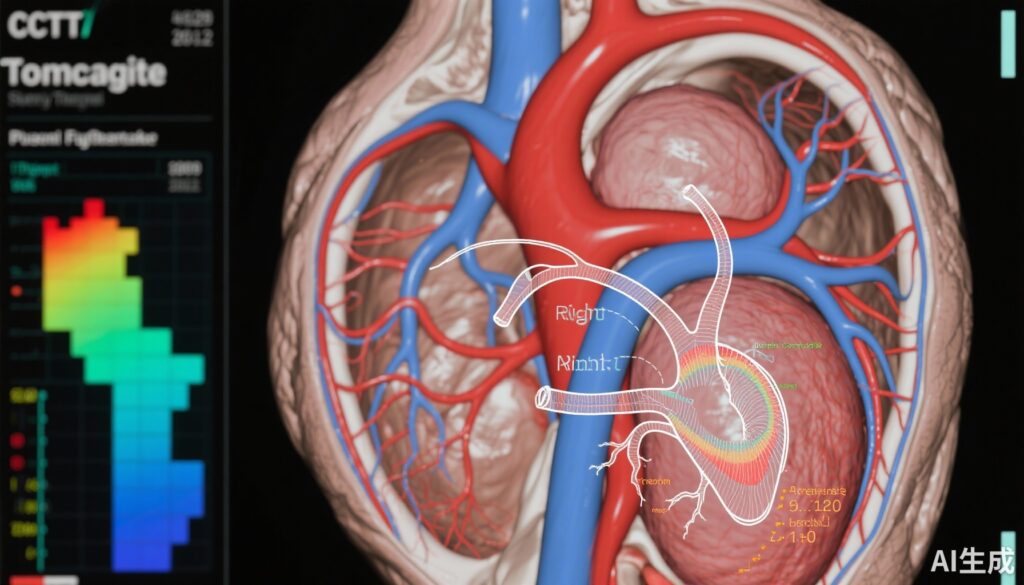
1. Coronary computed tomography angiography (CCTA) to define anatomical features, with particular focus on the ostial minor axis diameter.
2. Functional nuclear cardiac imaging to detect inducible ischemia.
3. Invasive fractional flow reserve measurement during dobutamine-atropine volume challenge (FFR-dobutamine), serving as the reference standard.
The primary endpoint was the determination of hemodynamic relevance, defined as an FFR-dobutamine value ≤0.8.
Key Findings
The study cohort included 55 patients with a mean age of 51 years (SD 12) and a predominance of males (67%). The median FFR-dobutamine was 0.87 (IQR 0.80-0.91), with 15 patients (27%) categorized as having hemodynamically relevant lesions (FFR ≤0.8).
Coronary Computed Tomography Angiography (CCTA) Performance
CCTA assessment using the ostial minor axis diameter demonstrated a remarkable diagnostic performance for excluding hemodynamic significance. It achieved:
– Sensitivity: 100%
– Negative Predictive Value (NPV): 100%
– Specificity: 57%
– Receiver Operating Characteristic Area Under Curve (ROC AUC): 0.82
Using CCTA, 23 cases (42%) were confidently ruled out as hemodynamically nonrelevant, correlating to 58% of all nonrelevant cases. This high sensitivity and NPV suggest that CCTA can effectively exclude patients from undergoing invasive FFR testing.
Functional Nuclear Imaging Performance
Functional nuclear imaging detected ischemia in 4 patients (7%), all of whom were true positives for hemodynamically relevant lesions per FFR-dobutamine. There were no false positives.
Diagnostic performance metrics included:
– Sensitivity: 27%
– Specificity: 100%
– Positive Predictive Value (PPV): 100%
– Accuracy: 80%
Though nuclear imaging exhibited low sensitivity, its high specificity and PPV imply it is valuable to confirm hemodynamic relevance when ischemia is observed.
Clinical Implications of Multimodality Diagnostic Approach
The findings support a stepwise diagnostic algorithm:
– Initial evaluation with CCTA to noninvasively and reliably exclude hemodynamically nonrelevant anatomical anomalies, reducing the need for invasive procedures.
– For patients with equivocal or suspicious findings on CCTA, adjunctive functional imaging can provide confirmatory evidence to rule in hemodynamic significance.
– Invasive FFR-dobutamine testing be reserved for patients with unresolved diagnostic ambiguity or where intervention is contemplated.
This approach balances diagnostic accuracy with patient safety, procedural resource utilization, and cost-effectiveness.
Expert Commentary
This study provides important evidence addressing a significant clinical challenge: noninvasive assessment of hemodynamic relevance in R-AAOCA. Previous reliance on invasive FFR for physiologic assessment limited accessibility and raised procedural risks. The demonstration of 100% sensitivity and NPV for anatomical CCTA endorsement as a rule-out tool is particularly reassuring.
However, limitations include the modest specificity of CCTA, underscoring that anatomical stenosis does not always equate to functional ischemia. Modest sensitivity of nuclear imaging reaffirms its role as a confirmatory but not standalone test. The exclusion of patients with atherosclerotic stenosis reduces confounding but may limit generalizability to mixed pathology populations.
Further multicenter studies with larger cohorts, integration of emerging imaging techniques like CT-derived FFR, and longitudinal outcome data are necessary to refine protocols and guidelines. Additionally, mechanistic insights into dynamic vessel compression during stress and intramural course impact on flow provide a strong pathophysiological rationale supporting these imaging strategies.
Conclusion
Right anomalous aortic origin of a coronary artery (R-AAOCA) poses a complex diagnostic challenge due to its potential for dynamic obstruction and ischemia. This prospective cohort study demonstrates the utility of a multimodality, stepwise, noninvasive imaging approach: initial anatomical evaluation with CCTA providing high sensitivity and negative predictive value to rule out hemodynamic relevance, and complementary functional nuclear imaging with high specificity to confirm ischemia. Such a strategy can substantially reduce reliance on invasive FFR-dobutamine testing, improving patient safety and optimizing resource use. These findings inform clinical algorithms, facilitating personalized diagnostic pathways for patients with R-AAOCA and addressing an important unmet need in congenital coronary anomaly care.
References
1. Bigler MR, Stark AW, Caobelli F, Rominger A, Kakizaki R, Biccirè FG, Al-Sabri SMA, Shiri I, Siepe M, Windecker S, Räber L, Gräni C. Noninvasive Anatomical and Functional Imaging for Hemodynamic Relevance in Right Coronary Artery Anomalies. JAMA Cardiol. 2025 Sep 10:e252993. doi: 10.1001/jamacardio.2025.2993. Epub ahead of print. PMID: 40928770; PMCID: PMC12423952.
2. Warnes CA, Williams RG, Bashore TM, et al. ACC/AHA 2008 Guidelines for the Management of Adults With Congenital Heart Disease. Circulation. 2008;118(23):e714-e833.
3. Angelini P. Coronary Artery Anomalies: A Comprehensive Approach. Lippincott Williams & Wilkins; 1999.
4. Petraco R, Park JM, Rana O, et al. Use of coronary computed tomography derived fractional flow reserve in patients with anomalous coronary arteries: A novel approach. J Cardiovasc Comput Tomogr. 2017;11(1):77-79.


