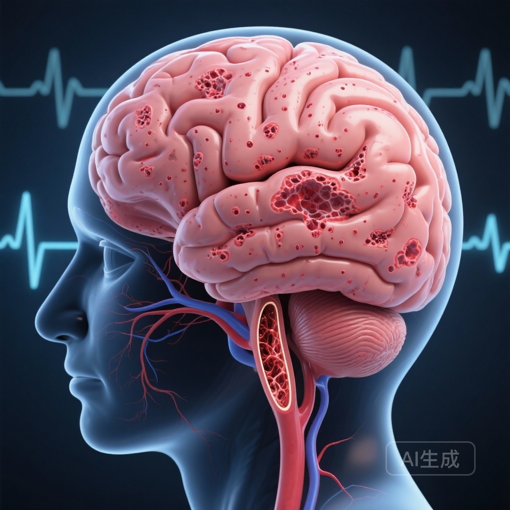
Introduction: The Subclinical Burden of Cardiovascular Disease
The relationship between cardiovascular health and brain integrity is one of the most critical frontiers in modern geriatric medicine and neurology. While the clinical manifestation of a stroke is a clear and devastating event, a much larger proportion of patients with cardiovascular disease harbor subclinical vascular brain lesions. These lesions, detectable primarily through magnetic resonance imaging (MRI), include silent brain infarcts, white matter hyperintensities (WMH), and cerebral micro-bleeds (CMB). These markers are not merely incidental findings; they are robust predictors of future clinical stroke, cognitive decline, and the development of dementia.
Historically, atrial fibrillation (AF) and atherosclerosis have been grouped together as primary drivers of cerebrovascular risk. However, the pathophysiological mechanisms by which they damage the brain are fundamentally different. AF is traditionally viewed as a source of cardioembolism, while atherosclerosis—particularly when affecting the carotid arteries or the intracranial vasculature—is associated with both embolic events and chronic hypoperfusion. Until recently, direct comparisons of the specific brain lesion patterns produced by these two distinct disease states were lacking. A landmark study published in the European Heart Journal (Stegmann et al., 2025) provides the first large-scale, standardized comparison of these neuroimaging signatures.
Study Design and Methodology: Leveraging the Swiss-AF and COMPASS MIND Cohorts
To elucidate the differences in lesion patterns, researchers conducted a cross-sectional analysis using baseline data from two major prospective studies: the Swiss Atrial Fibrillation cohort study (Swiss-AF) and the Cardiovascular Outcomes for People Using Anticoagulation Strategies (COMPASS) MRI sub-study (COMPASS MIND).
The combined analysis included 3,508 patients. The AF group (n = 1,748) was derived from Swiss-AF, representing a population with documented AF, the majority of whom (90%) were managed with oral anticoagulation. The atherosclerosis group (n = 1,760) consisted of patients from COMPASS MIND who had established coronary or peripheral artery disease but no history of AF. This group was primarily managed with anti-platelet therapy (93%).
The demographic profiles were remarkably similar, with a mean age of 73 in the AF cohort and 71 in the atherosclerosis cohort. The primary objective was to compare the prevalence and distribution of several key MRI markers: lacunar infarcts (small, deep lesions), non-lacunar infarcts (larger, often cortical lesions), periventricular and deep white matter hyperintensities (markers of small vessel disease), and cerebral micro-bleeds.
Key Findings: Divergent MRI Phenotypes
The study’s results underscore that AF and atherosclerosis are not interchangeable risk factors; they leave distinct footprints on the brain’s architecture.
Non-Lacunar vs. Lacunar Infarcts
Patients with atrial fibrillation were significantly more likely to exhibit non-lacunar infarcts compared to those with atherosclerosis (22% vs. 10%; P < .001). Conversely, patients with atherosclerosis showed a higher prevalence of lacunar infarcts (26% vs. 21%; P = .001). After adjusting for age, sex, and vascular risk factors in multivariable analyses, the odds of having non-lacunar infarcts were more than double in AF patients (OR 2.28; 95% CI 1.86–2.81). In contrast, the odds of lacunar infarcts were significantly lower in the AF group (OR 0.66; 95% CI 0.56–0.79) when compared to the atherosclerosis group.
White Matter Hyperintensities
White matter hyperintensities (WMH) are often used as a surrogate for cerebral small vessel disease. The study found that AF patients had a significantly higher grade of periventricular WMH (49% vs. 37%; P < .001). Even after multivariable adjustment, AF remained associated with higher odds of severe periventricular white matter disease (OR 1.42; 95% CI 1.22–1.67). Interestingly, the differences in deep white matter hyperintensities were less pronounced, suggesting that AF may specifically influence the periventricular vascular environment.
Cerebral Micro-bleeds (CMB)
Cerebral micro-bleeds are small deposits of iron from previous hemorrhages, often linked to hypertensive vasculopathy or cerebral amyloid angiopathy. These were more common in the atherosclerosis cohort (29%) than in the AF cohort (22%; P < .001). This suggests that the systemic burden of atherosclerosis may be more closely aligned with the small-vessel fragility that leads to micro-hemorrhages.
Mechanistic Insights: Embolism vs. Small Vessel Disease
The findings support the “disease-specific mechanism” hypothesis. The high prevalence of non-lacunar infarcts in AF patients—even those on anticoagulation—points to a persistent cardioembolic risk. These larger infarcts usually involve the cerebral cortex and are characteristic of emboli originating from the left atrium or auricle. The fact that many of these lesions are subclinical suggests that current anticoagulation strategies, while effective at preventing major strokes, may not fully eliminate the shower of smaller emboli that contribute to cumulative brain damage.
On the other hand, the preponderance of lacunar infarcts and micro-bleeds in the atherosclerosis group aligns with the classical understanding of systemic vascular disease. Atherosclerosis is frequently a systemic marker for cerebral small vessel disease (SVD), where lipohyalinosis and microatheroma of the small penetrating arteries lead to small, deep ischemic strokes (lacunes) and chronic ischemia (white matter changes).
The higher burden of periventricular WMH in AF patients is particularly intriguing. While WMH are typically associated with hypertension, some researchers suggest that the irregular cardiac output and fluctuating cerebral blood flow inherent in AF may lead to chronic hypoperfusion of the periventricular regions, which are particularly sensitive to changes in blood flow due to their “watershed” nature.
Expert Commentary: Clinical Implications and Future Directions
These findings have profound implications for how we manage patients with cardiovascular disease. For the clinician, an MRI showing multiple non-lacunar lesions in a patient without a diagnosis of AF should perhaps trigger more intensive cardiac monitoring. Conversely, a high burden of lacunar disease and micro-bleeds in a patient with AF might suggest that their risk profile is driven as much by systemic small vessel disease as by their arrhythmia, necessitating aggressive blood pressure and lipid management alongside anticoagulation.
However, the study is not without limitations. The cross-sectional nature prevents a definitive determination of causality. Furthermore, while the majority of AF patients were on anticoagulants, the impact of the duration and quality of anticoagulation on lesion development remains an area for further longitudinal study.
One of the most pressing questions raised by this data is the impact of these subclinical lesions on cognitive function. If AF leads to larger, more frequent (though silent) non-lacunar infarcts, does this explain the higher risk of dementia observed in AF patients, independent of clinical stroke? The Swiss-AF and COMPASS MIND cohorts are uniquely positioned to answer these questions as longitudinal data becomes available.
Conclusion: A Call for Personalized Cerebrovascular Care
The study by Stegmann et al. clarifies that atrial fibrillation and atherosclerosis are not merely general risk factors for “brain aging” but are drivers of specific, identifiable patterns of vascular injury. By recognizing that AF is strongly linked to non-lacunar infarcts and periventricular white matter disease, while atherosclerosis is linked to lacunar infarcts and micro-bleeds, physicians can better interpret neuroimaging findings and tailor preventive strategies to the specific underlying pathology. As we move toward a more personalized approach to medicine, understanding these distinct neuroimaging signatures will be essential in the fight against stroke and cognitive decline.
References
1. Stegmann T, Joundi RA, Srivastava A, et al. Atrial fibrillation and atherosclerosis cause different vascular brain lesions on magnetic resonance imaging. Eur Heart J. 2025;46(47):5177-5188. doi:10.1093/eurheartj/ehaf550.
2. Conen D, Rodondi N, Müller A, et al. Relationships of Atrial Fibrillation with Cognitive Decline and Dementia. Circulation. 2019;139(11):1421-1436.
3. Wardlaw JM, Smith EE, Biessels GJ, et al. Neuroimaging standards for research into small vessel disease and its contribution to ageing and neurodegeneration. Lancet Neurol. 2013;12(8):822-838.