Highlights
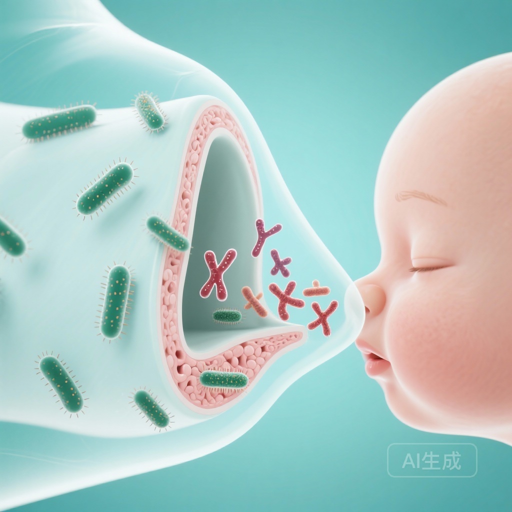
Maternal Tdap-IPV vaccination significantly increases nasal anti-pertussis toxin and anti-B. pertussis IgG in infants at 8 weeks of age, demonstrating effective transplacental transfer to the upper airway mucosa.
Infants receiving the whole-cell pertussis (DTwP) primary vaccine series developed significantly higher mucosal IgG and broader nasal T-cell cytokine responses compared to those receiving the acellular (DTaP) vaccine.
While maternal antibodies blunted the infant’s own pertussis toxin-specific IgG response, the superior mucosal activation of DTwP suggests a more robust defense against colonization and transmission.
Background: The Challenge of Pertussis Resurgence
Bordetella pertussis remains a significant cause of infant morbidity and mortality globally. Despite high vaccination coverage, many regions have witnessed a resurgence of the disease. A critical factor in this resurgence is the difference in the type of immunity conferred by acellular pertussis (DTaP) vaccines versus whole-cell pertussis (DTwP) vaccines. While both are effective at preventing severe clinical disease, DTaP vaccines appear less effective at preventing nasal colonization and transmission, likely due to a failure to induce robust mucosal resident memory T cells and local IgA responses.
To protect infants during the vulnerable first weeks of life before they complete their primary vaccine series, many countries have implemented maternal vaccination programs. However, the interaction between maternally derived antibodies and the infant’s subsequent response to different types of primary pertussis vaccines—particularly at the mucosal level—remains poorly understood. This study aims to bridge that gap by examining mucosal immunity in a clinical trial setting.
Study Design and Methodology
This immunological substudy was part of the Gambian Pertussis Study (GaPs), a single-centre, randomised, controlled, double-blind, phase 4 trial conducted in The Gambia. The study utilized a complex 2×2 factorial design to evaluate both maternal and infant vaccine interactions.
Population and Interventions
Healthy pregnant participants (aged 18–40 years) were randomized (1:1) to receive either a pertussis-containing vaccine (Tdap-IPV) or a tetanus-toxoid (TT) control vaccine between 28 and 34 weeks of gestation. Their infants were then randomized (1:1) to receive a primary immunization series of either DTwP or DTaP at 8, 12, and 16 weeks of age.
Sample Collection and Endpoints
The researchers used nasosorption devices to collect nasal mucosal lining fluid (MLF) at 8, 16, 17, 20 weeks, and 9 months of age. Paired cord blood and serum samples were also analyzed. The primary outcomes for this substudy included the concentrations of nasal anti-B. pertussis IgG and IgA, anti-pertussis toxin (PT) IgG, and a panel of T-cell-associated cytokines (including IL-17 and IFN-gamma) to assess local cellular activation.
Key Findings: Maternal Transfer and Infant Response
The substudy included 160 infants, providing a detailed look at the kinetics of mucosal immunity from birth through the first nine months of life.
Maternal Antibody Transfer to the Mucosa
At 8 weeks of age, before the infants received their first primary dose, those born to mothers who received Tdap-IPV had significantly higher concentrations of nasal IgG compared to the control group. The geometric mean ratio (GMR) for anti-PT IgG was 3.84 (95% CI 3.22–4.59; p<0.0001), and for anti-B. pertussis IgG, it was 6.45 (95% CI 5.94–7.01; p<0.0001). Notably, no significant difference was found in nasal IgA, suggesting that the protection provided by maternal vaccination is primarily IgG-mediated and transplacentally derived.
Superiority of DTwP in Inducing Mucosal Immunity
Following the primary vaccination series, the DTwP vaccine demonstrated a clear advantage in inducing local immunity. At 20 weeks, infants in the DTwP groups had significantly higher nasal anti-B. pertussis IgG concentrations than those in the DTaP group. For example, the TT-DTwP group reached 5.42 AU/mL compared to only 2.16 AU/mL in the Tdap-IPV-DTaP group.
T-Cell Cytokine Activation
Perhaps the most striking finding was the difference in cellular responses. At 17 weeks, infants vaccinated with DTwP showed broad nasal T-cell-associated cytokine responses, including elevations in IFN-gamma and IL-17, which are critical for clearance of B. pertussis from the airway. In contrast, these responses were largely absent in infants who received the DTaP vaccine. This provides clinical evidence for the DTwP-specific mucosal effects previously identified only in animal models.
The Blunting Effect
The study confirmed the phenomenon of “immunological blunting.” Infants whose mothers received Tdap-IPV had lower anti-PT IgG responses after their own vaccinations compared to infants whose mothers received TT. This blunting was observed in both the DTwP and DTaP groups but was more pronounced in the DTwP group (GMC 0.016 IU/mL vs. 0.073 IU/mL). Despite this reduction in specific PT-IgG, the DTwP infants still maintained higher overall mucosal IgG to the whole bacterium than DTaP infants.
Expert Commentary and Clinical Implications
The results of this substudy offer a mechanistic explanation for why whole-cell vaccines are associated with better herd immunity and lower transmission rates. The induction of a Th1/Th17 cytokine profile in the nasal mucosa by DTwP likely facilitates the recruitment and activation of mucosal-resident memory T cells, which are the first line of defense against colonization.
From a policy perspective, these findings support the continued use of DTwP in regions where it is currently standard, while highlighting the limitations of DTaP in controlling the spread of the bacteria. The evidence that maternal antibodies reach the infant’s upper respiratory tract reinforces the value of antenatal immunization as a strategy to protect newborns before they are old enough to be vaccinated.
Study Limitations
While the study provides robust immunological data, it was a single-centre study in The Gambia, which may affect generalizability to populations with different nutritional statuses or environmental exposures. Additionally, while mucosal antibodies and cytokines were measured, the study did not directly measure the prevention of B. pertussis colonization in these infants.
Conclusion
This phase 4 substudy demonstrates that maternal Tdap-IPV vaccination effectively populates the infant’s nasal mucosa with protective IgG. Furthermore, it establishes that the infant primary series with DTwP induces a more comprehensive mucosal immune profile—characterized by higher IgG levels and significant T-cell activation—than the DTaP vaccine. These findings are crucial for informing future vaccine design and optimizing global immunization schedules to achieve better control of pertussis transmission.
Funding and ClinicalTrials.gov
This study was funded by the Innovative Medicines Initiative 2 Joint Undertaking, Horizon 2020, European Federation of Pharmaceutical Industries and Associations, the Bill & Melinda Gates Foundation, Wellcome Trust, and BactiVac. It is registered with ClinicalTrials.gov, NCT03606096.
References
1. Saso A, Fröberg J, Jobe H, et al. Mucosal immune responses to Bordetella pertussis in Gambian infants after maternal and primary vaccination: an immunological substudy of a single-centre, randomised, controlled, double-blind, phase 4 trial. Lancet Microbe. 2026 Jan;7(1):101219. doi: 10.1016/lanmic.2025.101219.
2. Kilgore PE, Magder LS, Weiner LB, et al. Pertussis in Infants: The Role of Maternal Antibody and Infant Vaccination. Journal of Infectious Diseases. 2016;213(11):1753-1761.
3. Wilk MM, Misiak A, McManus RM, et al. Cyclophosphamide and IL-1R Antagonist Reduce the Efficacy of Whole-Cell Pertussis Vaccine by Affecting Th17 Cells and Mucosal Resident Memory T Cells. Frontiers in Immunology. 2019;10:1759.