Patient Information
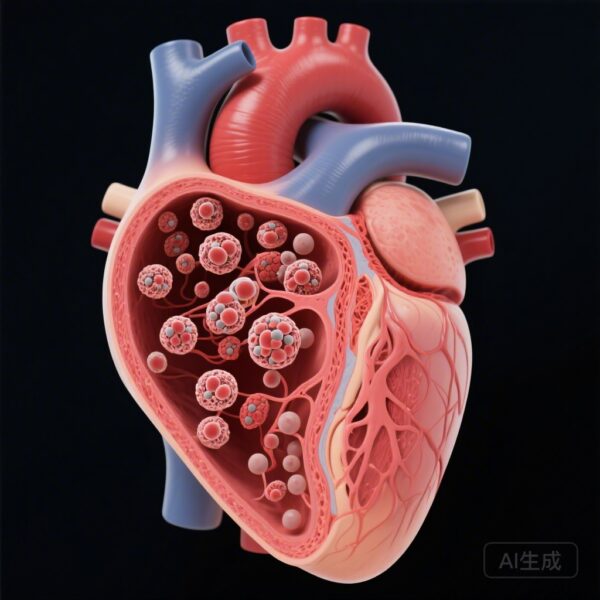
Coronary artery ectasia (CAE) is defined as a localized or diffuse dilation of the coronary artery lumen that exceeds the diameter of adjacent normal segments by at least 1.5 times. While CAE often occurs in the presence of atherosclerotic coronary artery disease, ‘isolated’ CAE refers to those cases occurring without significant obstructive stenosis. In patients presenting with their first acute myocardial infarction (MI), isolated CAE represents a distinct clinical entity. Demographically, patients in this cohort often present with symptoms of acute coronary syndrome, though the underlying pathophysiology differs from standard plaque rupture. In the study population of 404 patients analyzed, a majority presented with ST-elevation myocardial infarction (STEMI), indicating a total or near-total acute occlusion of the ectatic vessel.
Diagnosis
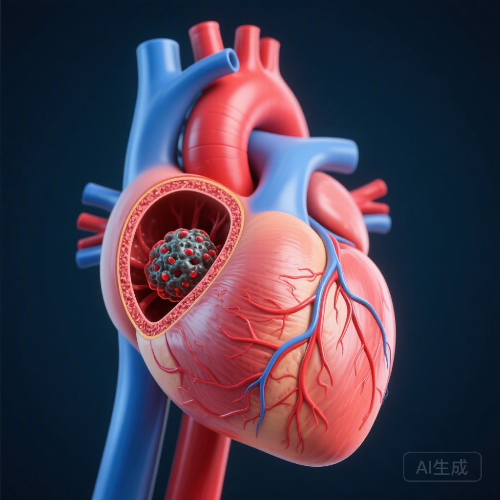
The diagnosis of isolated CAE is primarily established through coronary angiography. Key findings in these cases include marked luminal dilation in the absence of >20% stenosis in any other vessel. Diagnosis is further refined using the Markis classification system, which categorizes the extent of ectasia. In patients with MI due to isolated CAE, the distribution is frequently diffuse and multivessel. Specifically, data indicates that approximately 71.3% of these patients are classified as Markis I (diffuse ectasia in two or three vessels) and 26.7% as Markis II (diffuse ectasia in one vessel and localized ectasia in another). The reasoning for the MI diagnosis in these patients involves the identification of a culprit lesion within an ectatic segment, where sluggish blood flow (slow-flow phenomenon) and blood stasis predispose the patient to thrombus formation.
Differential Diagnosis
When a patient presents with MI, several conditions must be ruled out alongside or instead of isolated CAE:
- Obstructive Atherosclerotic Coronary Artery Disease (CAD): The most common cause of MI, ruled out here by the absence of significant (>20%) stenosis.
- Coronary Artery Aneurysm: Distinguished from ectasia by being a localized dilation rather than a diffuse one.
- Vasospastic Angina (Prinzmetal’s): Ruled out by the anatomical findings on angiography.
- Spontaneous Coronary Artery Dissection (SCAD): Identified by the presence of an intimal tear or intramural hematoma on imaging.
- Myocardial Bridge: A congenital variant where a segment of a coronary artery is covered by myocardium, usually ruled out via angiography.
Treatment and Management
The management of MI in the setting of isolated CAE is complex. Standard primary percutaneous coronary intervention (PCI) is often the first-line treatment for STEMI. However, the large caliber of ectatic vessels presents unique challenges, including difficulty in sizing stents and an increased risk of ‘no-reflow’ due to the high thrombus burden. In the study group, patients received standard-of-care medical therapy, yet the incidence of stent thrombosis was significantly higher (8.9% in the CAE group versus 1.5% in the control group). This suggests that the decision-making process for stenting in dilated vessels must be approached with caution. Adjunctive pharmacotherapy, including intensive antiplatelet therapy and potentially long-term anticoagulation, is often considered due to the inherent stasis of blood within the ectatic segments, though specific guidelines for isolated CAE are still evolving.
Outcome and Prognosis
The long-term prognosis for patients with MI caused by isolated CAE is notably poorer than for those with MI but without CAE. Over the follow-up period, the composite outcome of all-cause death and nonfatal recurrent MI occurred in 26.7% of the isolated CAE group compared to 16.3% in the control group (P=0.011).
Recurrent MI was a significant driver of this disparity, occurring in 22.8% of CAE patients versus 13.4% of controls. Multiple variable analysis confirmed that the presence of CAE was an independent risk factor, with a hazard ratio (HR) of 1.84 for death/recurrent MI and a HR of 2.07 for recurrent MI specifically. Furthermore, the rate of recurrent MI originating from the original infarct artery was significantly higher in the CAE cohort, likely due to the persistent pro-thrombotic environment of the dilated vessel.
Discussion
This case report and associated study data underscore the clinical significance of isolated diffuse coronary artery ectasia as more than just a morphological curiosity. The findings suggest that isolated CAE is a high-risk condition when it leads to myocardial infarction. The unique pathophysiology—characterized by blood flow stasis and a propensity for thrombus—leads to a higher rate of both spontaneous recurrent MI and mechanical complications like stent thrombosis.
One of the most critical lessons learned is the high rate of stent thrombosis (8.9%), which is nearly six times higher than in patients with non-ectatic vessels. This suggests that the traditional approach to stenting may need modification in CAE patients, perhaps favoring balloon angioplasty, thrombus aspiration, or aggressive anticoagulation over drug-eluting stents in extremely large vessels where apposition is poor.
Clinicians should view isolated CAE as a chronic systemic vascular condition requiring rigorous secondary prevention and potentially more aggressive antithrombotic strategies. Further research is required to determine if long-term oral anticoagulation can mitigate the high recurrence rates observed in this population.
References
1. Abaci A, Yerlikaya MG, et al. Long-Term Prognosis of Acute Myocardial Infarction Caused by Isolated Diffuse Coronary Artery Ectasia. Circ Cardiovasc Interv. 2026. doi: 10.1161/CIRCINTERVENTIONS.125.016071.
2. Markis JE, Joffe CD, et al. Clinical significance of coronary arterial ectasia. Am J Cardiol. 1976;37(2):217-22.
3. Devabhaktuni S, et al. Coronary Artery Ectasia – A Review. Southern Medical Journal. 2016;109(9):530-539.