Background: The Evolution of Cardiac Resynchronization Therapy
Cardiac resynchronization therapy (CRT) has long been established as a cornerstone treatment for patients with dyssynchronous heart failure (DHF), a condition characterized by abnormal electrical conduction that causes inefficient ventricular contraction and progressive cardiac dysfunction. Traditional biventricular pacing (BiVP), which delivers electrical impulses to both ventricles simultaneously, has demonstrated significant clinical benefits including improved symptoms, reduced hospitalizations, and enhanced survival in appropriately selected patients.
However, BiVP is not without limitations. The technique requires placement of leads in both the right ventricle and coronary sinus tributaries, procedures that can be technically challenging and associated with complications including coronary sinus dissection, phrenic nerve stimulation, and suboptimal lead positioning. Furthermore, despite advances in device technology and patient selection criteria, approximately 30% of patients fail to respond adequately to BiVP-CRT, underscoring the need for alternative pacing strategies that might achieve superior physiological activation patterns.
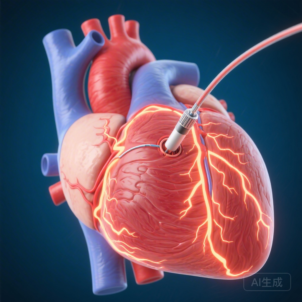
Left bundle branch pacing (LBBP) has emerged as a promising alternative approach that directly targets the conduction system by positioning the pacing lead distal to the His-Purkinje network. This strategy aims to achieve more physiological ventricular activation than conventional BiVP, potentially resulting in better mechanical synchrony and reverse remodeling. The study by Huang and colleagues, published in the European Heart Journal, provides mechanistic insights comparing these two pacing modalities using a comprehensive canine model of dyssynchronous heart failure.
Study Design and Methodology
The investigators employed a well-characterized canine model to explore the differential effects of LBBP versus BiVP on cardiac structure, function, and molecular signaling. The study enrolled 31 adult canines, which were allocated into four experimental groups following left bundle branch ablation to establish the dyssynchronous heart failure substrate.
The dyssynchronous heart failure group (DHF, n=8) underwent six weeks of rapid atrial tachypacing at 200 beats per minute to induce heart failure. The biventricular pacing group (BiVP, n=8) received three weeks of atrial tachypacing followed by three weeks of simultaneous BiVP during continued tachypacing. The left bundle branch pacing group (LBBP, n=8) underwent an identical protocol with LBBP replacing BiVP during the final three weeks. A non-intervened control group (n=7) served as normal reference.
Comprehensive assessments were performed including surface electrocardiography to measure QRS duration, echocardiography to evaluate left ventricular ejection fraction (LVEF) and global longitudinal strain (LVGLS), and molecular analyses to quantify biomarkers of heart failure, cytoskeleton proteins, transforming growth factor-beta (TGF-β) signaling pathways, sarcoplasmic reticulum calcium ATPase 2a (SERCA2a) expression, and myocardial energy metabolism markers.
Key Electrocardiographic and Echocardiographic Findings
Both pacing modalities achieved substantial and statistically equivalent reductions in QRS duration from baseline dyssynchronous values. The BiVP-treated canines demonstrated QRS narrowing of 32 ± 6 milliseconds, while LBBP-treated animals showed 35 ± 5 milliseconds reduction (P = .276). This equivalence indicates that both approaches successfully normalized ventricular activation times, achieving the primary electrophysiological goal of cardiac resynchronization.
Regarding global systolic function, both treatment arms produced meaningful improvements in left ventricular ejection fraction. BiVP increased LVEF by 9 ± 5 percentage points, and LBBP achieved a comparable 10 ± 4 percentage point improvement (P = .390). These equivalent gains in LVEF suggest that both pacing strategies effectively reverse the severe systolic dysfunction induced by dyssynchronous pacing.
However, the most striking difference emerged when examining left ventricular global longitudinal strain, a more sensitive marker of myocardial deformation and subclinical ventricular function. LBBP demonstrated a significantly greater improvement in LVGLS compared to BiVP, with changes of -3.7 ± 1.2% versus -2.3 ± 1.0% respectively (P = .019). The more negative strain values indicate superior restoration of longitudinal myocardial shortening, suggesting that LBBP achieves more physiological contraction patterns that more closely approximate normal cardiac mechanics.
Molecular and Cellular Insights
Beyond conventional imaging parameters, the study conducted extensive molecular profiling that revealed fundamental differences in how the two pacing strategies influence cardiac biology at the cellular and subcellular levels.
Both BiVP and LBBP effectively reversed circulating and tissue biomarkers of heart failure, indicating successful suppression of the neurohormonal activation and inflammatory cascade characteristic of decompensated heart failure. However, the magnitude and breadth of molecular recovery differed substantially between groups.
LBBP demonstrated significantly more pronounced modulation of cytoskeleton proteins compared to BiVP. Cytoskeletal remodeling is a hallmark of pathological cardiac hypertrophy and failure, and the enhanced normalization of these structural proteins with LBBP suggests superior reversal of maladaptive structural changes at the sarcomeric and cellular levels.
The TGF-β signaling pathway, a central mediator of cardiac fibrosis and adverse remodeling, showed greater suppression with LBBP than with BiVP. This finding implies that LBBP may confer additional benefits in preventing or reversing interstitial fibrosis, potentially contributing to improved diastolic function and long-term cardiac outcomes beyond the study period.
Perhaps most importantly, SERCA2a expression was more significantly upregulated with LBBP compared to BiVP. SERCA2a is the critical calcium handling protein responsible for reuptake of calcium into the sarcoplasmic reticulum during diastole, and its downregulation is a fundamental abnormality in heart failure that contributes to both systolic and diastolic dysfunction. The superior restoration of SERCA2a with LBBP indicates more complete recovery of calcium cycling, which underlies improved excitation-contraction coupling and cardiac performance.
Myocardial Energy Metabolism
The study also provided compelling evidence that LBBP contributes to more comprehensive improvement in myocardial energy metabolism compared to BiVP. Heart failure is characterized by a metabolic shift from efficient fatty acid oxidation toward less efficient glucose metabolism, along with mitochondrial dysfunction and ATP depletion. The differential effects of pacing modality on metabolic recovery have important implications for understanding why some patients respond better to physiological activation patterns.
The enhanced metabolic recovery observed with LBBP likely reflects the more physiological nature of ventricular activation achieved through direct Purkinje system stimulation. When electrical impulses propagate through the native conduction system rather than being delivered directly to myocardium, the pattern of calcium release and contraction is more coordinated, potentially reducing wasted energy from dyssynchronous work and improving overall cardiac efficiency.
Mechanistic Implications
These findings provide important mechanistic insights into why LBBP might offer advantages over traditional BiVP in clinical practice. The conduction system originates in the sinus node and spreads through the atrioventricular node, bundle of His, and Purkinje network to achieve rapid, coordinated ventricular activation. BiVP, while effective, delivers electrical impulses to ventricular myocardium from arbitrary locations, which can result in activation fronts meeting at arbitrary collision points and creating regions of mechanical dyssynchrony even when QRS duration appears normalized.
LBBP, by capturing the left bundle branch distal to the site of conduction block, leverages the intact Purkinje network to achieve activation patterns that more closely approximate normal physiology. The result is more homogeneous contraction, improved systolic mechanics as evidenced by superior LVGLS improvement, and better preservation of the delicate molecular machinery governing calcium handling and energy metabolism.
The superior outcomes in TGF-β signaling and cytoskeleton protein modulation with LBBP suggest that physiological activation may provide not just symptomatic improvement but actual disease modification by preventing progressive structural deterioration. These molecular signatures of reverse remodeling could translate into more durable clinical benefits and improved long-term prognosis compared to conventional resynchronization approaches.
Expert Commentary and Clinical Perspectives
While these preclinical findings are compelling, several important considerations must be addressed before drawing definitive clinical conclusions. The canine model, while providing valuable mechanistic insights, differs from human heart failure in important ways including etiology, comorbidities, and response to chronic disease. The acute induction of dyssynchronity through bundle branch ablation followed by rapid pacing may not fully recapitulate the chronic, progressive nature of human cardiomyopathy.
The three-week treatment duration, while substantial for preclinical studies, may not capture the full spectrum of adaptive and maladaptive responses that develop over months to years of pacing therapy in clinical practice. Long-term studies will be essential to determine whether the molecular advantages of LBBP translate into sustained functional improvement and survival benefits.
Furthermore, the technical aspects of LBBP implantation in humans present unique challenges including the need for precise lead positioning within or near the left bundle branch, potential for perforation or injury to the conduction system, and learning curve considerations for implanting physicians. While early clinical series have demonstrated feasibility and safety in experienced centers, widespread adoption will require standardized implantation techniques and comprehensive operator training.
Nevertheless, the mechanistic superiority of LBBP across multiple domains—electrical, mechanical, and molecular—provides a strong rationale for continued clinical investigation. The findings support the physiological plausibility that direct conduction system pacing may offer advantages over conventional approaches, and justify randomized clinical trials comparing these strategies in patients with guideline-indicated CRT.
Conclusion
This comprehensive canine study demonstrates that left bundle branch pacing achieves cardiac resynchronization effects at least equivalent to traditional biventricular pacing when measuring traditional electrocardiographic and functional parameters. Both modalities similarly narrow QRS duration and improve left ventricular ejection fraction. However, LBBP demonstrates clear superiority in more sensitive assessments of myocardial mechanics, achieving significantly greater improvement in global longitudinal strain.
The mechanistic insights from molecular analyses reveal that LBBP provides more comprehensive recovery at the cellular and subcellular levels, with enhanced normalization of cytoskeleton proteins, TGF-β signaling, SERCA2a expression, and myocardial energy metabolism. These findings suggest that physiological activation through the native conduction system may offer disease-modifying benefits beyond simple electrical resynchronization.
While these preclinical observations require validation in human clinical trials, they provide compelling evidence that left bundle branch pacing represents a promising evolution in cardiac resynchronization therapy. As implant techniques continue to refine and clinical experience accumulates, LBBP may emerge as a preferred strategy for patients with dyssynchronous heart failure who require permanent pacing support.
Funding and Study Registration
This research was supported by institutional grants as detailed in the original publication. The study protocol received appropriate institutional animal care and use committee approval, and all procedures were conducted in accordance with established ethical standards for animal research.
References
Huang H, Hu Y, Li H, Cheng S, Jiang Y, Weng S, He P, Yang J, Chen G, Liu X, Pei J, Chen L, Gu M, Ding L, Niu H, Jin H, Pan X, Vernooy K, Prinzen FW, Hua W. Left bundle branch vs biventricular pacing: mechanistic insights from a canine model. Eur Heart J. 2026 Apr 1;47(13):1595-1605. PMID: 41528069.