Highlights
1. Reduced lung function, characterized by lower FEV1 and FVC, is significantly associated with systemic insulin resistance and impaired insulin clearance across the diabetes spectrum.
2. Mendelian randomization (MR) analysis provides evidence for a causal relationship between HOMA-insulin resistance (HOMA-IR) and decreased FEV1.
3. Patients with Type 2 diabetes, particularly those with severe insulin resistance (SIRD), exhibit more pronounced pulmonary deficits than those with Type 1 diabetes or healthy controls.
4. Higher daily insulin doses in Type 1 diabetes are inversely correlated with lung function, suggesting that hyperinsulinemia may exert direct adverse effects on pulmonary tissue.
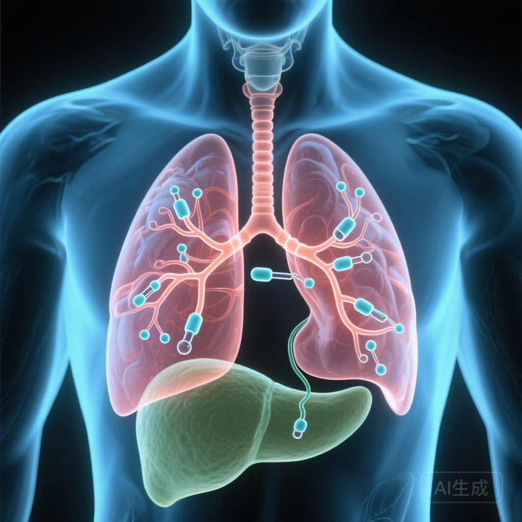
Introduction: The Lung as a Forgotten Target of Diabetes
For decades, clinical focus in diabetes management has prioritized the prevention of microvascular and macrovascular complications affecting the kidneys, retina, nerves, and cardiovascular system. However, emerging evidence suggests that the lung may be another “target organ” of diabetes. While clinical observations have long linked diabetes to reduced forced expiratory volume in 1 second (FEV1) and forced vital capacity (FVC), the underlying mechanisms—whether they are driven by hyperglycemia, systemic inflammation, or specific insulin-related metabolic pathways—have remained elusive. The study by Di Marco et al., published in Diabetes Care (2026), provides a pivotal analysis of how metabolic factors modulate the connection between diabetes and pulmonary alterations, utilizing both cross-sectional data from the German Diabetes Study (GDS) and causal inference via Mendelian randomization.
Study Design and Methodological Rigor
The research employed a dual-approach methodology to ensure both clinical depth and statistical causality. First, the investigators analyzed 1,152 participants from the German Diabetes Study (GDS), comprising 426 individuals with Type 1 diabetes, 482 with Type 2 diabetes, and 244 healthy controls (CON). A defining strength of the GDS cohort is the use of high-precision phenotyping, including Botnia clamps to measure insulin sensitivity (M-value), insulin secretion, and insulin clearance.
To move beyond correlation and address potential confounding, the researchers conducted Mendelian randomization (MR) analysis. This technique uses genetic variants as instrumental variables to assess the causal effect of metabolic traits on lung function. Data for the MR were sourced from the MAGIC (Meta-Analyses of Glucose and Insulin-Related Traits Consortium) for insulin resistance (n = 37,037) and the UK Household Longitudinal Study for pulmonary parameters (n = 321,047).
Results: Quantifying the Impact of Insulin Sensitivity on Lung Mechanics
The findings from the GDS cohort revealed a clear metabolic gradient in pulmonary health. Higher insulin sensitivity (M-value) and more efficient insulin clearance were consistently associated with higher FEV1 and FVC (all β > 0.18, P < 0.0001). Conversely, participants with Type 2 diabetes demonstrated significantly lower lung function parameters compared to both Type 1 diabetes patients and healthy controls. This deficit was most pronounced in specific diabetes subgroups, namely those with severe insulin resistance (SIRD) and those with mild age-related diabetes (MARD).
Insulin Dosing and Pulmonary Function
Interestingly, in patients with Type 1 diabetes, FEV1 was negatively associated with daily insulin doses (β = -0.21, P = 0.0006). This suggests that exogenous hyperinsulinemia, often required to overcome peripheral resistance or manage glycemic volatility, may contribute to the remodeling or stiffening of pulmonary tissue. In the Type 2 diabetes cohort, FEV1 was specifically lower in the SIRD cluster (β = -0.27, P = 0.039), further reinforcing the hypothesis that the severity of insulin resistance is a primary determinant of lung impairment.
Causality via Mendelian Randomization
The MR analysis provided the definitive “smoking gun” for the metabolic hypothesis. The results supported a causal association between HOMA-IR and lower FEV1 (β = -0.13, P = 0.0018). Importantly, the study found that this relationship was not merely a byproduct of obesity or general systemic inflammation, but rather a direct consequence of insulin-related signaling pathways.
Expert Commentary: Mechanistic Insights and Clinical Implications
The association between insulin resistance and pulmonary decline likely involves several intersecting pathways. From a physiological standpoint, the lung is a highly vascularized organ with a massive basement membrane surface area. Chronic hyperinsulinemia is known to stimulate the proliferation of airway smooth muscle cells and increase the deposition of collagen in the lung parenchyma. These structural changes, akin to the microvascular thickening seen in diabetic nephropathy, lead to reduced lung compliance and restricted airflow.
Furthermore, insulin resistance is often accompanied by a state of chronic low-grade inflammation. However, the MR data suggesting a causal link independent of BMI indicates that insulin signaling itself—or the lack of effective signaling—plays a direct role. Insulin is involved in the production of pulmonary surfactant and the maintenance of alveolar epithelial integrity. When this system is disrupted, the lung’s resilience to environmental stressors or aging is diminished.
Clinical Considerations
For clinicians, these findings suggest that pulmonary function tests (PFTs) might be undervalued in the routine assessment of patients with severe insulin resistance. While most diabetic patients do not present with overt respiratory failure, the subclinical reduction in FEV1 and FVC can limit exercise tolerance, thereby exacerbating the metabolic cycle of inactivity and weight gain. Furthermore, in patients with Type 1 diabetes, the negative correlation with high insulin doses should prompt a re-evaluation of strategies to improve insulin sensitivity (such as adjunctive metformin or SGLT2 inhibitors in selected cases) to potentially mitigate pulmonary side effects.
Conclusion: Integrating Pulmonary Health into Diabetes Care
The study by Di Marco and colleagues marks a significant step forward in our understanding of the multi-organ impact of diabetes. By identifying insulin resistance and impaired clearance as causal factors in pulmonary decline, the research elevates the lung from an “innocent bystander” to a key participant in the systemic pathology of diabetes. Future research should investigate whether aggressive insulin-sensitizing therapies can halt or reverse these pulmonary alterations, and whether lung function can serve as a non-invasive biomarker for overall metabolic health.
References
1. Di Marco M, Bódis KB, Li Z, et al. Metabolic Factors Modulating the Connection Between Diabetes and Pulmonary Alterations. Diabetes Care. 2026;49(3):426-434. PMID: 41474454.
2. Klein OL, Krishnan JA, Glick S, Smith LJ. Systematic review: the association between lung function and type 2 diabetes mellitus. Diabetologia. 2010;53(12):2479-2490.
3. Pitocco D, Fuso L, Conte EG, et al. The diabetic lung: a new target organ? Reviews on Diabetic Studies. 2012;9(1):23-35.
4. Rawshani A, Rawshani A, Franzén S, et al. Risk Factors, Mortality, and Cardiovascular Outcomes in Patients with Type 2 Diabetes. N Engl J Med. 2018;379:633-644.