The Hidden Landscape of Coronary Risk: Insights into Silent Plaque Ruptures
Highlights
- Approximately 12% of patients presenting with acute myocardial infarction (AMI) harbor silent plaque ruptures in non-obstructive lesions of non-infarct-related arteries.
- Ruptured plaques in non-infarct-related arteries (non-IRAs) are characterized by significantly larger plaque volumes, positive remodeling, and thinner fibrous caps compared to non-ruptured lesions.
- More than half of these untreated silent ruptures transition into stable, healed morphologies within 52 weeks of follow-up.
- Thin-cap fibroatheroma (TCFA) is the most frequent baseline morphology for new-onset silent ruptures, reinforcing its role as a precursor to clinical and subclinical events.
Introduction: The Systemic Nature of Acute Coronary Syndromes
For decades, the management of acute myocardial infarction (AMI) has focused heavily on the identification and treatment of the culprit lesion—the specific site of plaque rupture or erosion that leads to acute vessel occlusion. However, clinical experience and autopsy studies have long suggested that atherosclerosis is not merely a focal disease but a systemic inflammatory process. Patients with AMI often possess multiple vulnerable plaques throughout the coronary tree, a phenomenon sometimes referred to as pan-coronary vulnerability.
While obstructive lesions are the primary targets for revascularization, non-obstructive lesions often remain under the clinical radar. Recent advancements in multimodality intracoronary imaging have allowed clinicians to look beyond the culprit lesion to understand the biological behavior of these non-obstructive plaques. A critical question has remained: how often do these non-obstructive plaques rupture without causing symptoms, and what is their natural history? The study by Kakizaki et al., published in the European Heart Journal, provides definitive insights into these “silent” plaque ruptures.
Study Design: Leveraging IBIS-4 and PACMAN-AMI Data
To investigate the prevalence and evolution of silent plaque ruptures, researchers conducted a serial, multimodality imaging analysis using pooled data from two major clinical trials: IBIS-4 and PACMAN-AMI. This study population consisted of patients who presented with AMI and underwent successful percutaneous coronary intervention (PCI) of the culprit lesion.
The unique aspect of this research was the systematic imaging of non-infarct-related arteries (non-IRAs). Patients underwent high-resolution imaging at baseline (during the index AMI event) and again at a 52-week follow-up. The imaging protocol utilized three distinct modalities to provide a comprehensive view of plaque morphology:
1. Intravascular Ultrasound (IVUS): Used to assess plaque volume and vessel remodeling.
2. Optical Coherence Tomography (OCT): Provided near-histological resolution to measure fibrous cap thickness and identify ruptures.
3. Near-Infrared Spectroscopy (NIRS): Employed to quantify the lipid content of the plaques.
By analyzing 783 lesions from 336 patients, the researchers were able to track the morphological life cycle of both existing and new-onset silent ruptures.
Key Findings: The Prevalence and Anatomy of Silent Rupture
The study found that silent plaque ruptures in non-obstructive lesions of non-IRAs were surprisingly common, occurring in 12% of AMI patients (41 lesions in 40 patients). These ruptures occurred in lesions that did not significantly impede blood flow, explaining why they did not manifest as secondary ischemic events at the time of the initial presentation.
Morphological Predictors of Rupture
When comparing lesions with silent ruptures to those without, several key morphological differences emerged. Lesions that had undergone silent rupture exhibited:
– Larger Percent Atheroma Volume (PAV): 53.3% vs. 49.5%, indicating a higher overall burden of disease within the vessel wall.
– Positive Remodeling: These lesions showed a larger external elastic membrane (EEM) area (20.5 mm2 vs. 15.7 mm2). Positive remodeling is a process where the vessel wall expands outward to accommodate plaque growth, maintaining the lumen size but often resulting in a more unstable plaque structure.
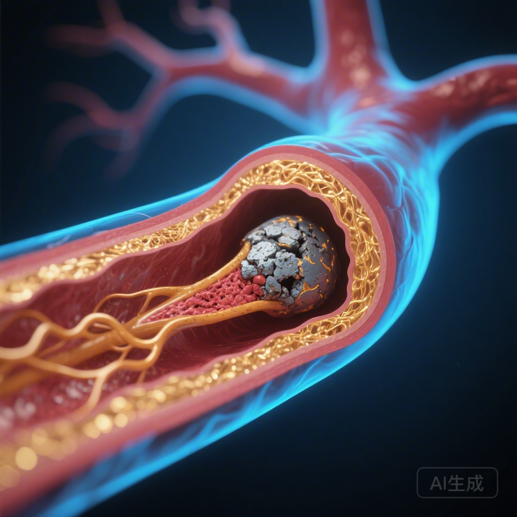
– Thinner Fibrous Caps: The minimum fibrous cap thickness (FCT) was significantly lower in ruptured lesions (69 μm vs. 116 μm). This aligns with the established threshold of 65 μm often used to define thin-cap fibroatheroma (TCFA), the most vulnerable type of plaque.
Interestingly, systemic biomarkers—including levels of circulating lipids and inflammatory markers—were comparable between patients with and without silent ruptures. This suggests that the occurrence of these ruptures may be driven more by local mechanical and morphological factors than by systemic laboratory values alone.
The Fate of Silent Ruptures: Healing and Transformation
One of the most clinically significant aspects of this study was the 52-week follow-up. Of the 41 silent rupture sites identified at baseline, 21 (51%) were found to have “healed” by the end of the year. In these cases, the imaging showed the restoration of a continuous fibrous cap over the previous rupture site, effectively stabilizing the lesion.
This healing process is a testament to the dynamic nature of atherosclerosis. It suggests that many plaque ruptures occur subclinically and resolve through the body’s endogenous repair mechanisms, often aided by intensive medical therapy (such as high-intensity statins and other lipid-lowering agents used in the trial protocols).
However, the study also identified 10 new-onset silent ruptures at the follow-up imaging that were not present at baseline. The most frequent precursor for these new ruptures was TCFA. This finding reinforces the concept that the “vulnerable plaque” is a moving target; while some lesions stabilize, others progress to rupture, even in the absence of acute symptoms.
Expert Commentary: Clinical Implications and Future Directions
The findings from Kakizaki et al. have profound implications for how we view coronary artery disease. First, they confirm that the presence of a culprit lesion in one artery is often a marker for widespread instability across the entire coronary tree. The 12% prevalence of silent ruptures in non-IRAs highlights the need for a shift from a “lesion-centric” approach to a “patient-centric” approach in secondary prevention.
The Role of Medical Therapy
In both the IBIS-4 and PACMAN-AMI trials, patients received intensive lipid-lowering therapy. The fact that more than half of the silent ruptures healed suggests that aggressive medical management is effective in promoting plaque stabilization. This supports current guidelines that advocate for achieving very low LDL-C targets in post-AMI patients to not only prevent new events but also to facilitate the healing of existing subclinical injuries.
Limitations and Considerations
While the study provides high-quality serial imaging data, it is important to note its limitations. The sample size, while robust for an imaging study, is relatively small compared to large-scale clinical outcome trials. Additionally, the study focused on AMI patients; the prevalence and behavior of silent ruptures in patients with stable coronary artery disease may differ. Furthermore, while imaging can identify a “healed” appearance, the long-term mechanical strength of these healed caps compared to original stable plaques remains a subject of investigation.
Conclusion: A Dynamic Paradigm of Risk
This multimodality imaging study provides a clear window into the silent progression of coronary atherosclerosis. Silent plaque ruptures are frequent in the wake of an AMI, occurring in one out of every eight patients in non-obstructive, non-culprit lesions. These events are driven by high plaque volume and thin fibrous caps, yet they show a remarkable capacity for healing under modern medical management.
For clinicians, the takeaway is clear: the absence of symptoms or significant stenosis in a non-infarct-related artery does not equate to the absence of risk. The high rate of TCFA-to-rupture transition emphasizes that we must remain vigilant in our systemic treatment of AMI patients. The goal of therapy is not just to fix the lesion that caused the heart attack, but to stabilize the entire coronary environment, preventing the next silent rupture from becoming a loud clinical catastrophe.
References
1. Kakizaki R, Biccirè FG, Losdat S, et al. Silent plaque ruptures in non-obstructive lesions of non-infarct-related arteries: a multimodality, serial intracoronary imaging study. European Heart Journal. 2026. PMID: 41795942.
2. Räber L, Ueki Y, Otsuka T, et al. Effect of Alirocumab Added to High-Intensity Statin Therapy on LDL Cholesterol Levels and Plaque Burden in Patients With Acute Myocardial Infarction: The PACMAN-AMI Randomized Clinical Trial. JAMA. 2022;327(18):1771–1781.
3. Windecker S, Haude M, Neumann FJ, et al. 2018 ESC/EACTS Guidelines on myocardial revascularization. European Heart Journal. 2019;40(2):87-165.