Highlights
1. Annexin A2 (ANXA2) is significantly upregulated in human and murine ischemic hearts, and its circulating levels correlate with the severity of acute myocardial infarction (MI).
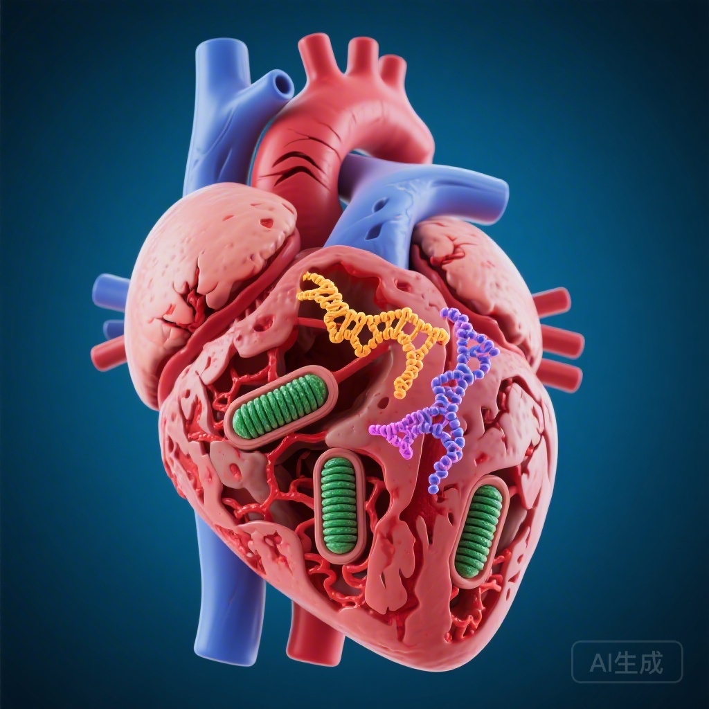
2. ANXA2 acts as a molecular brake on mitophagy by competitively binding to the receptor Prohibitin 2 (PHB2), preventing its interaction with LC3B.
3. Mechanistically, ANXA2 recruits the E3 ligase TRIM29 to mediate the K48-linked polyubiquitination and proteasomal degradation of PHB2.
4. Cardiomyocyte-specific depletion of ANXA2 restores mitochondrial quality control, reduces oxidative stress, and significantly improves cardiac function and remodeling post-MI.
Background: The Challenge of Mitophagy in Ischemic Heart Disease
Myocardial infarction remains a leading cause of heart failure and mortality worldwide. Following an ischemic event, the heart undergoes profound structural and functional changes, collectively known as cardiac remodeling. Central to this process is the health of the cardiomyocyte mitochondrial pool. Mitophagy—the selective autophagic degradation of damaged mitochondria—is a vital quality control mechanism that maintains cellular homeostasis under stress. While basal mitophagy is protective, its dysregulation during and after ischemia-reperfusion or permanent infarction can exacerbate cell death and fibrotic remodeling.
The Annexin A family of proteins is known to participate in membrane trafficking and calcium-dependent signaling, but its role in orchestrating mitophagy within the infarcted myocardium has remained elusive. Specifically, Annexin A2 (ANXA2) has been implicated in various inflammatory and fibrinolytic processes, yet its intrinsic influence on mitochondrial survival pathways in cardiomyocytes represents a critical gap in our understanding of post-MI pathophysiology.
Study Design: A Multi-Omics and Translational Approach
To investigate the role of ANXA2, the research team employed a comprehensive array of experimental models and techniques. Transcriptome analysis was initially used to identify differentially expressed genes in infarcted murine hearts. The study then transitioned to clinical validation, examining ANXA2 expression in human ischemic failing hearts and measuring circulating ANXA2 levels in patients with acute MI.
The functional impact of ANXA2 was tested using cardiomyocyte-specific Anxa2 knockdown (AAV9-shAnxa2) and overexpression models in mice subjected to permanent left anterior descending (LAD) artery ligation. These in vivo findings were complemented by in vitro studies using neonatal rat ventricular myocytes (NRVMs) exposed to hypoxia. To elucidate the molecular mechanism, the researchers utilized immunoprecipitation (IP), mass spectrometry (MS), and glutathione S-transferase (GST) pull-down assays to map the interactome of ANXA2, eventually focusing on its relationship with the mitophagy receptor Prohibitin 2 (PHB2) and the E3 ligase TRIM29.
Key Findings: ANXA2 as a Pathological Driver of Mitophagy Failure
Clinical Correlation and Expression Patterns
The researchers observed that ANXA2 expression was markedly elevated in the border zones of infarcted hearts in both mice and humans. Furthermore, in patients presenting with acute MI, plasma levels of ANXA2 were positively correlated with markers of cardiac injury, such as troponin levels, and inversely correlated with left ventricular ejection fraction (LVEF). This suggested that ANXA2 might not just be a marker of injury but a participant in the pathological process.
ANXA2 Depletion Ameliorates Cardiac Injury
In the murine MI model, cardiomyocyte-specific depletion of Anxa2 led to a significant preservation of cardiac function. Compared to control MI mice, those with Anxa2 knockdown exhibited reduced infarct size, decreased cardiomyocyte apoptosis, and attenuated inflammatory cell infiltration. Echocardiographic parameters showed improved LVEF and reduced ventricular dilation. Conversely, overexpression of Anxa2 exacerbated mitochondrial fragmentation, increased oxidative stress, and worsened heart failure outcomes, establishing ANXA2 as a detrimental factor in the post-MI environment.
Restoration of Mitophagy
A pivotal finding was that ANXA2 levels inversely regulated the rate of mitophagy. Under hypoxic conditions, ANXA2 was found to translocate or increase its association with mitochondria. Knocking down Anxa2 prevented the decline in mitophagy typically seen in the later stages of ischemic stress, thereby facilitating the clearance of dysfunctional, ROS-producing mitochondria. This effect was shown to be dependent on the presence of the mitophagy receptor PHB2.
Mechanistic Insights: The ANXA2-TRIM29-PHB2 Axis
The study provides a detailed molecular map of how ANXA2 suppresses mitophagy. PHB2 is an inner mitochondrial membrane protein that, upon mitochondrial depolarization or damage, becomes accessible to the autophagosomal protein LC3B, thereby bridging the mitochondrion to the autophagosome.
Competitive Inhibition
The researchers discovered that ANXA2 directly interacts with the LC3-interacting region (LIR) of PHB2. By binding to this site, ANXA2 competitively blocks LC3B from recognizing damaged mitochondria, effectively halting the mitophagic process at its recognition phase.
Ubiquitination and Degradation
Beyond competitive inhibition, ANXA2 acts as a scaffold to facilitate the degradation of the PHB2 protein itself. The study identified TRIM29 (tripartite motif-containing 29) as the specific E3 ligase recruited by ANXA2 to the mitochondrial surface. ANXA2 promotes TRIM29-mediated K48-linked polyubiquitination of PHB2, leading to its proteasomal degradation. This double-hit mechanism—blocking the receptor and then destroying it—explains the potent inhibitory effect of ANXA2 on mitochondrial quality control.
Clinical Significance and Expert Commentary
The identification of the ANXA2-PHB2 axis represents a significant shift in our understanding of how the heart manages mitochondrial stress. While much research has focused on the PINK1/Parkin pathway, this study highlights the importance of receptor-mediated mitophagy (via PHB2) and its post-translational regulation.
From a clinical perspective, ANXA2 presents a dual-purpose target. First, as a biomarker, circulating ANXA2 could potentially aid in the risk stratification of post-MI patients. Second, pharmacological inhibition of ANXA2—or the disruption of the ANXA2-PHB2 interaction—could serve as a novel therapeutic strategy to limit remodeling. However, experts note that Annexin A2 has multifaceted roles in other tissues, including blood coagulation and fibrinolysis. Therefore, any future therapeutic intervention would ideally need to be targeted to the myocardium to avoid systemic side effects, such as bleeding diathesis.
The study also raises interesting questions regarding the timing of mitophagy. While the authors demonstrate that inhibiting ANXA2 is beneficial in the acute and subacute phases of MI, the long-term effects of sustained mitophagy activation require further longitudinal investigation to ensure that ‘over-clearing’ of mitochondria does not eventually lead to metabolic deficits.
Conclusion
In summary, the research by Deng et al. elucidates a sophisticated regulatory mechanism where ANXA2 serves as a molecular ‘brake’ on mitophagy in the wake of myocardial infarction. By orchestrating the degradation of PHB2 via TRIM29 and physically shielding the PHB2-LC3B interaction, ANXA2 prevents the necessary removal of damaged mitochondria, leading to increased oxidative stress and cardiac dysfunction. The finding that Anxa2 deficiency restores mitochondrial homeostasis and improves functional recovery offers a compelling new lead in the quest to treat ischemic heart disease and prevent the progression to heart failure.
References
1. Deng KQ, Xu Z, Wang Q, et al. Inhibition of Annexin A2 Facilitates PHB2-Mediated Mitophagy in Cardiomyocytes to Alleviate Cardiac Injury and Remodeling After Infarction. Circulation. 2026;153(11):826-844. PMID: 41492949.
2. Yan X, et al. Mitochondrial Quality Control in Myocardial Infarction: Mechanisms and Therapeutic Targets. Nature Reviews Cardiology. 2023.
3. Wei Y, et al. Prohibitin 2 Interacts with LC3 and Functions as an Inner Mitochondrial Membrane Mitophagy Receptor. Cell. 2017;168(1-2):224-238.