Introduction: The Evolving Landscape of Hepatocellular Carcinoma Research
Hepatocellular carcinoma (HCC) remains one of the most lethal malignancies worldwide, characterized by high recurrence rates and limited therapeutic options for advanced-stage patients. While traditional genomic and transcriptomic studies have identified several key drivers of hepatocarcinogenesis, the functional complexity of metabolic enzymes in cancer progression is only beginning to be understood. Among these, aminoacyl-tRNA synthetases (ARSs), traditionally known for their canonical role in protein translation, are emerging as multifaceted regulators of cellular homeostasis and oncogenic transformation.
A groundbreaking study published in Hepatology (2026) by Hermán-Sánchez and colleagues has shed light on Aspartyl-tRNA synthetase 1 (DARS1), revealing it as a pivotal player in HCC. The research demonstrates that DARS1 does not merely facilitate protein synthesis but actively reshapes the HCC proteome and drives aggressiveness through a non-canonical nuclear mechanism involving the SAGA-MYC signaling axis.
Highlights of the Research
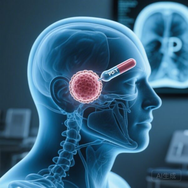
– DARS1 is significantly overexpressed in HCC tissues and detectable in patient plasma, marking its potential as a non-invasive liquid biopsy biomarker.
– Beyond its cytoplasmic role in translation, DARS1 translocates to the nucleus, where it interacts with the SAGA transcriptional co-activator complex.
– DARS1 modulation directly affects MYC stability and activity; its depletion induces senescence and impairs cancer stemness.
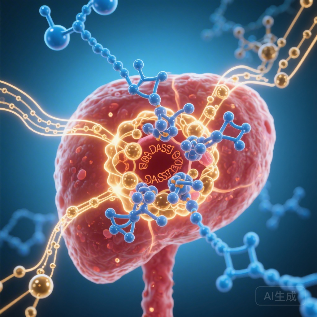
– The study identifies a unique “proteome reshaping” effect, where DARS1 overexpression specifically enriches the abundance of high-aspartate proteins involved in tumor progression.
Background: The Unmet Need in HCC and the Role of Aspartate Metabolism
Aspartate metabolism is frequently altered in HCC to meet the high biosynthetic demands of rapidly proliferating cells. As the enzyme responsible for charging tRNA with aspartate, DARS1 is at the heart of this metabolic reprogramming. However, the extent to which DARS1 contributes to HCC pathogenesis—beyond its basic metabolic function—has remained largely unexplored until now. Given the clinical challenge of identifying reliable biomarkers and therapeutic targets for aggressive HCC subtypes, investigating the clinical and functional significance of DARS1 represents a critical step forward.
Study Design and Methodology
The researchers employed a comprehensive, multi-omics approach to evaluate DARS1. The study utilized:
Clinical Validation
Analysis of DARS1 expression in matched tumor and non-tumoral tissues from HCC patients, supplemented by large-scale in silico cohorts (e.g., TCGA) and plasma samples for biomarker assessment.
Functional Characterization
In vitro assays using various liver cancer cell lines were conducted following the genetic knockdown (siRNA/shRNA) or pharmacological modulation of DARS1. The team specifically looked at parameters of aggressiveness, including cell proliferation, migration, invasion, and cancer stemness.
In Vivo Models
The oncogenic potential of DARS1 was validated using both subcutaneous xenograft and orthotopic liver tumor models in mice.
Mechanistic Insights
Quantitative proteomics (TMT-labeling) and immunoprecipitation-mass spectrometry (IP-MS) were used to identify DARS1-interacting proteins. Biochemical assays, including nuclear/cytoplasmic fractionation and luciferase reporter assays, were performed to elucidate the interaction between DARS1, the SAGA complex, and MYC signaling.
Key Findings: A Dual Mechanism of Oncogenesis
1. DARS1 as a Clinical and Diagnostic Indicator
The study found that DARS1 is consistently upregulated in HCC compared to healthy liver tissue. Notably, higher DARS1 levels correlated with increased tumor grade, advanced stage, and poorer patient prognosis. Perhaps most significantly for clinical practice, DARS1 was found to be significantly elevated in the plasma of HCC patients, suggesting its utility as a liquid biopsy marker for early detection or monitoring of treatment response.
2. Reshaping the HCC Proteome
Proteomic profiling revealed that DARS1 overexpression does not lead to a global, uniform increase in all proteins. Instead, it causes a selective enrichment of proteins with high aspartate content. Many of these proteins are involved in critical oncogenic pathways, suggesting that DARS1 selectively “tunes” the proteome to favor a more aggressive phenotype.
3. The Non-Canonical Nuclear SAGA-MYC Axis
The most striking discovery was the localization of DARS1 within the nucleus. The researchers identified an interaction between DARS1 and members of the SAGA (Spt-Ada-Gcn5 acetyltransferase) transcriptional co-activator complex, specifically SUPT7L. This interaction is crucial for the regulation of MYC, a master oncogene.
The study demonstrated that DARS1 depletion leads to:
– Reduced MYC protein levels.
– Increased MYC phosphorylation (associated with degradation).
– Enhanced drug-induced senescence in cancer cells.
By stabilizing MYC through the SAGA complex, DARS1 maintains the proliferative and self-renewal capacity of HCC cells, particularly the cancer stem cell (CSC) population.
4. Therapeutic Vulnerability
Pharmacological and genetic blockade of DARS1 effectively impaired HCC cell stemness and reduced tumor growth in vivo. Importantly, these interventions appeared to spare “normal-like” hepatocytes, suggesting a therapeutic window that minimizes off-target toxicity.
Expert Commentary and Mechanistic Insights
This study adds to a growing body of evidence that aminoacyl-tRNA synthetases are not just “housekeeping” enzymes but are active participants in cellular signaling. The dual role of DARS1—facilitating the translation of aspartate-rich proteins while simultaneously acting as a transcriptional modulator in the nucleus—provides a sophisticated model of how cancer cells coordinate metabolism and gene expression.
From a clinical perspective, the finding that DARS1 levels in plasma mirror tissue expression is highly promising. While alpha-fetoprotein (AFP) remains the standard biomarker for HCC, its sensitivity and specificity are often suboptimal. DARS1 could potentially complement AFP in diagnostic panels. Furthermore, the link between DARS1 and MYC is particularly relevant, as MYC itself is notoriously difficult to target directly. By targeting DARS1, clinicians may be able to indirectly dampen MYC-driven oncogenesis.
Conclusion and Future Directions
The study by Hermán-Sánchez et al. identifies DARS1 as a novel vulnerability in hepatocellular carcinoma. Its role in reshaping the proteome and modulating the SAGA-MYC axis highlights the complexity of metabolic enzymes in cancer. As we move toward more personalized medicine, DARS1 stands out as both a potential diagnostic tool and a target for innovative therapeutic strategies aimed at disrupting the aggressive backbone of HCC.
Future research should focus on developing high-affinity small-molecule inhibitors of DARS1 that can specifically disrupt its nuclear interactions without compromising essential systemic translation, as well as validating DARS1’s performance in larger, prospective liquid biopsy trials.
References
1. Hermán-Sánchez N, Fernández-Barrena MG, Alañón-Martínez PE, et al. Aspartyl-tRNA synthetase 1 (DARS1) reshapes hepatocellular carcinoma proteome and promotes aggressiveness through non-canonical SAGA-MYC signalling modulation. Hepatology. 2026. PMID: 41790991.
2. Kim S, Plotnikov AN, Kyratsous CA, et al. Functional diversification of aminoacyl-tRNA synthetases and their roles in cancer. Nature Reviews Cancer. 2011;11(10):708-718.
3. Sun A, Jiang J, Wang X, et al. Metabolic reprogramming in hepatocellular carcinoma: Mechanisms and clinical implications. Journal of Hematology & Oncology. 2020;13(1):161.