Highlights
• In a Medicare claims analysis (Jan 1, 2022–Dec 31, 2023), 19,692 patients underwent balloon sinus dilation (BSD); 15% (2,905) lacked a documented preprocedure CT within one year.
• Of 490 otolaryngologists included, 156 (31.8%) were identified as outliers performing BSD without a recent CT in >10% of their cases; 30 physicians accounted for >50% of missed CTs.
• While most clinicians followed consensus practice, concentrated nonadherence suggests targeted quality-improvement and monitoring interventions are warranted.
Background
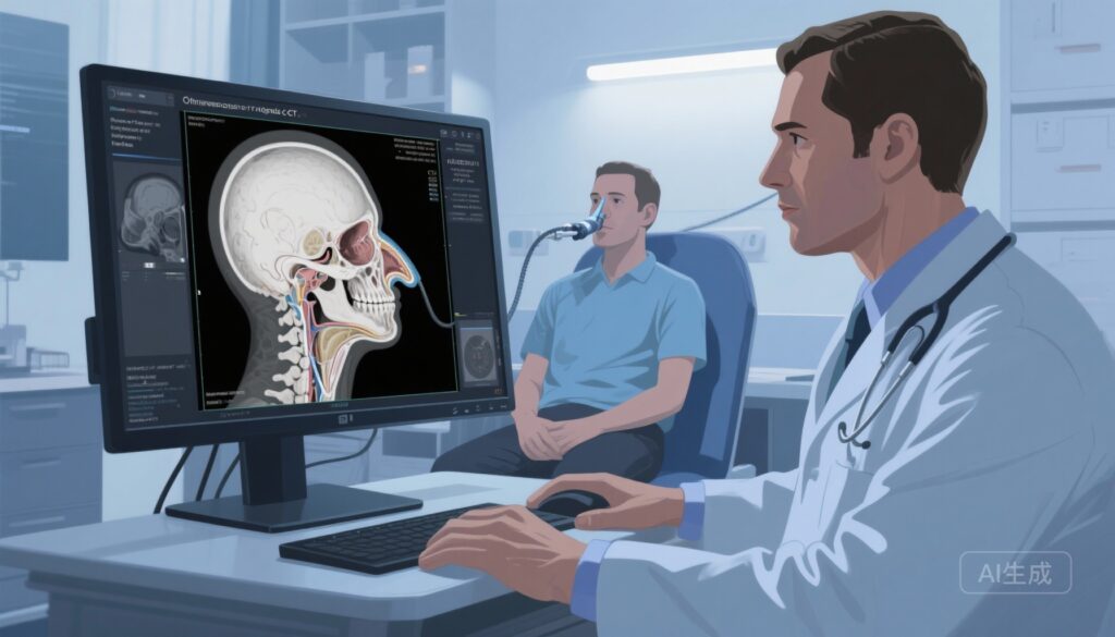
Balloon sinus dilation (BSD) is an established, minimally invasive intervention for selected patients with chronic rhinosinusitis and recurrent acute rhinosinusitis. Preoperative computed tomography (CT) of the paranasal sinuses is widely accepted as a necessary step in preoperative planning for sinus procedures because CT provides definitive anatomic delineation, identifies anatomic variants and disease extent, and guides both patient selection and operative strategy. Clinical practice guidelines and international consensus statements (for example, AAO‑HNSF and EPOS) recognize imaging as integral to decision-making for surgical therapy of the sinuses.
Study design
The study by Romashko et al. (JAMA Otolaryngol Head Neck Surg. 2025) is a cross-sectional analysis of Medicare claims used to evaluate adherence to guideline-concordant use of preoperative CT imaging before BSD. Inclusion criteria required otolaryngologists to have performed at least 11 BSDs during the two-year window (Jan 1, 2022–Dec 31, 2023). Imaging availability was defined as presence of CT imaging within 1 year prior to the BSD procedure. The primary outcome was the percentage of physicians who performed BSDs without CT imaging in >10% of their procedures (designated as outliers). Secondary analyses explored physician-level variables associated with outlier status. Data were analyzed using IBM-SPSS Statistics version 29 and R 4.3.2.
Key findings and detailed results
Population and scope. The analysis included 490 otolaryngologists (29 female [5.9%], 460 male [94.1%]) and 19,692 patients who underwent BSD. The study focused on a high-volume operator cohort, excluding surgeons performing fewer than 11 BSDs during the study period.
Overall imaging rates. Preprocedure CT imaging was not available within 1 year for 2,905 patients, representing 15.0% of the cohort. Thus, 85% of BSD procedures had a documented CT within the year preceding the procedure.
Provider-level distribution and outliers. Of the 490 clinicians, 156 (31.8%) met the study’s definition of outliers — that is, they performed BSD without a CT available within 1 year in more than 10% of their procedures. Strikingly, 30 otolaryngologists (6.1% of the sample) accounted for over 50% of cases without preprocedure CT (1,880 patients, or 9.5% of the total). In addition, 42 clinicians (8.6%) accounted for 47.5% of all missed CTs.
Concentration of nonadherence. These findings show a concentrated pattern: most clinicians obtained preoperative CTs routinely, while a minority of providers were responsible for a disproportionate share of the lapses. This concentration has two practical implications—targeted interventions at a relatively small number of practices could remediate a large fraction of nonadherent cases, and aggregated claims-level surveillance could identify outlier clinicians for quality review.
Interpretation of the 1-year window. The authors selected a 1-year lookback to capture cases where prior CTs might reasonably inform current surgical planning. This window balances recency with pragmatic intervals for chronic conditions; however, shorter or longer windows would change absolute counts, and the clinical relevance of older imaging depends on disease dynamics and prior interventions.
Subgroup and sensitivity considerations. The published abstract and summary emphasize the overall percentages and distribution of outliers. Claims-based analyses do not include granular clinical detail such as symptom severity, polyp status, prior imaging performed outside Medicare billing, in-office cone-beam CT availability, or surgeon judgment about when imaging is unnecessary (for example, recent imaging from other payers or radiology systems unavailable in claims). These limitations affect interpretation but do not negate the core finding of substantial provider-level variation.
Expert commentary and contextualization
Clinical standards and safety considerations. Both the AAO‑HNSF clinical practice guideline for adult sinusitis (Rosenfeld et al., 2015) and the European Position Paper on Rhinosinusitis and Nasal Polyps (EPOS 2020) underscore the role of cross-sectional imaging (CT) in surgical planning. CT imaging identifies critical anatomic variants (e.g., dehiscent lamina papyracea, Onodi cells, sphenoid septations, anterior ethmoidal artery location) that, if unrecognized, increase risk of complications. For clinicians, a preoperative CT is a tool for optimizing patient selection and minimizing perioperative risk.
Possible explanations for observed nonadherence. Several hypotheses could explain why some BSD procedures occurred without a documented CT in Medicare claims: (1) imaging performed but billed to a different insurer or performed outside the Medicare billing system (e.g., private imaging center, VA, or self-pay) and thus not captured; (2) very recent previous CTs just outside the selected lookback window; (3) in-office imaging such as cone-beam CT that was not billed to Medicare in a way captured by the dataset; (4) clinical judgment that CT was unnecessary in select cases, although this is not typical practice; or (5) documentation/billing errors. Claims data cannot categorically distinguish these scenarios.
Quality, policy and medico-legal implications. The concentration of missed imaging among a small group of providers raises quality and patient safety flags. Absent clear clinical justification, omission of preoperative CT may expose patients to avoidable risk and clinicians to medicolegal vulnerability. For payers and regulators, claims surveillance offers a pragmatic approach to monitor adherence to practice standards. For professional societies, these findings could prompt renewed emphasis on education, documentation standards, and consensus clarifications about acceptable exceptions.
Limitations of the study. Key limitations relate to the use of administrative claims: lack of clinical granular data (symptoms, endoscopic findings, prior imaging outside claims), potential misclassification if imaging was done but not captured in the dataset, inability to link CT findings to outcomes (complication rates, symptom improvement), and selection bias by restricting to higher-volume operators. The study therefore identifies variation and potential gaps, but cannot quantify clinical harm from missing imaging.
Clinical and policy recommendations
1. Reinforce guideline-consistent practice: Otolaryngology departments and professional societies should reiterate the role of CT in preoperative planning for BSD and provide decision aids clarifying rare, acceptable exceptions.
2. Targeted quality improvement: Because a small number of clinicians accounted for a large share of missed CTs, targeted audit-and-feedback interventions at the practice level could be efficient. Peer comparison reports, chart- or claims-based reminders, and local workflow changes (e.g., mandatory preoperative imaging checks) may reduce lapses.
3. Claims-based monitoring: Payers and health systems can deploy claims surveillance to identify outliers; such monitoring should, however, be coupled with mechanisms to adjudicate legitimate exceptions and to account for imaging billed through other channels.
4. Research priorities: Future work should link imaging utilization to clinical outcomes (perioperative complications, revision surgery, patient-reported outcomes) and examine reasons for missing imaging through chart reviews or prospective registries. Studies should also evaluate the role of in-office cone-beam CT and how its documentation intersects with claims datasets.
Conclusion
The Medicare claims analysis by Romashko et al. documents that while the majority of otolaryngologists obtain preoperative CT imaging before BSD, a clinically meaningful minority of procedures (15%) lacked a documented CT within one year, and this nonadherence was concentrated among a small subset of providers. Given the established value of imaging for safe and effective sinus surgery, these results underscore opportunities for targeted quality-improvement actions, clearer documentation practices, and further research to link imaging use to outcomes. Claims-based surveillance can identify outliers, but clinical validation is needed to inform proportionate responses.
Funding and clinicaltrials.gov
This study used Medicare claims data and is observational in design. Funding details and disclosures are those reported in the original manuscript; the design does not involve a clinical trial registration. Readers should consult the original publication for specific funding statements and disclosures.
References
1. Romashko AA, Farrell NF, Kallogjeri D, Sáenz MP, Smith KA, Piccirillo JF. Preoperative Computed Tomography Utilization in Patients Undergoing Balloon Sinus Dilation. JAMA Otolaryngol Head Neck Surg. 2025 Nov 13:e254030. doi: 10.1001/jamaoto.2025.4030. Epub ahead of print. PMID: 41231495; PMCID: PMC12616526.
2. Rosenfeld RM, Piccirillo JF, Chandrasekhar SS, et al. Clinical practice guideline (update): adult sinusitis. Otolaryngol Head Neck Surg. 2015 Jan;152(2 Suppl):S1-S39. doi:10.1177/0194599814561600.
3. Fokkens WJ, Lund VJ, Hopkins C, et al. European Position Paper on Rhinosinusitis and Nasal Polyps 2020. Rhinology. 2020 Feb;58(Suppl S29):1-464.