Introduction: The Neurologic Vulnerability of Children on ECMO
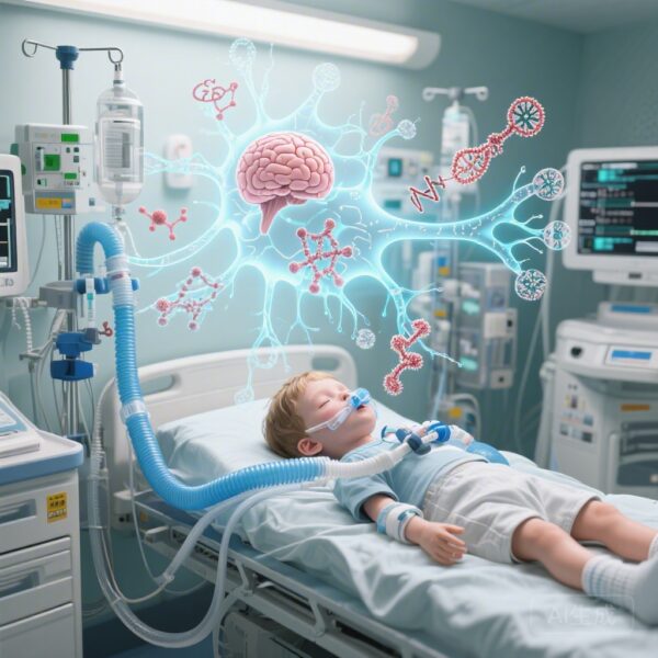
Extracorporeal membrane oxygenation (ECMO) is a life-saving intervention for children with refractory cardiac or respiratory failure. However, the procedure carries a high risk of acute brain injury (ABI), including ischemic stroke, intracranial hemorrhage, and hypoxic-ischemic encephalopathy. Identifying these injuries in real-time is notoriously difficult. Children on ECMO are often sedated, paralyzed, and clinically unstable, making traditional bedside neurologic examinations unreliable and transport to neuroimaging suites (CT or MRI) extremely hazardous.
There is an urgent clinical need for non-invasive, bedside tools that can detect brain injury as it occurs. A recent study by Friedman et al., published in JAMA Pediatrics, investigates whether plasma biomarkers—specifically glial fibrillary acidic protein (GFAP), neurofilament light chain (NfL), and tau—can serve as early warning signals for ABI and predictors of long-term functional outcomes in this high-risk population.
Study Design and Methodology
This prospective observational cohort study, conducted by the Pediatric Acute Lung Injury and Sepsis Investigators (PALISI) Network, recruited 219 children (representing 224 ECMO courses) across 11 US children’s hospitals between 2019 and 2023. The study population included neonates as young as two days old up to adolescents under 18 years.
Researchers collected serial blood samples during the ECMO course to measure concentrations of GFAP (a marker of glial/astrocyte injury), NfL (a marker of axonal damage), and tau (a marker of microtubule-associated axonal and somatic injury). The primary objectives were to determine if these biomarkers rose before ABI was confirmed on neuroimaging and if they correlated with short-term and long-term outcomes. Long-term outcomes were assessed at 18 months post-ECMO using the Vineland Adaptive Behavior Scales, third edition.
Key Findings: Biomarkers as Early Warning Signals
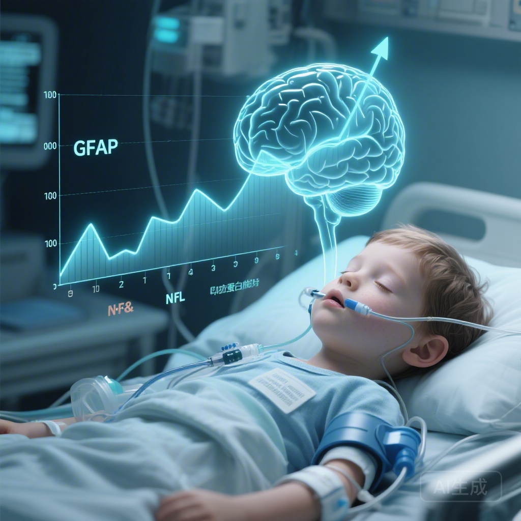
Among the 224 ECMO courses, 60 children developed a new ABI confirmed by neuroimaging. The study found that plasma levels of GFAP and NfL began to rise significantly well before the injury was visible on scans.
Preceding the Diagnosis
For every 24 hours preceding a neuroimaging-confirmed diagnosis of ABI, GFAP levels increased by 6.4% (95% CI, 1.4%-11.6%) and NfL levels increased by 16.1% (95% CI, 10.5%-22.0%). This suggests that these proteins leak into the bloodstream as soon as the cellular integrity of the brain is compromised, providing a potential window for clinicians to intervene before the injury progresses or becomes permanent.
Association with Mortality and Functional Decline
At hospital discharge, geometric means for GFAP, NfL, and tau were significantly higher in children with unfavorable outcomes (mortality or significant decline in Pediatric Cerebral Performance Category scores).
The study highlighted the prognostic power of GFAP and NfL kinetics. After adjusting for age, baseline levels, and the reason for ECMO, a two-fold increase in GFAP from the initial sample was associated with a 48% higher risk of unfavorable outcomes (adjusted Hazard Ratio [aHR], 1.48; 95% CI, 1.22-1.79). Similarly, a two-fold increase in NfL was associated with a 43% higher risk (aHR, 1.43; 95% CI, 1.14-1.79). Interestingly, while tau levels were elevated in some cases, they did not show a statistically significant association with long-term outcomes in the adjusted models.
Expert Commentary: A Paradigm Shift in Neuro-Monitoring
The findings from this PALISI Network study represent a significant step toward precision neuro-monitoring in the pediatric intensive care unit. The ability of GFAP and NfL to predict ABI before imaging is particularly transformative.
GFAP is highly specific to the central nervous system and is released following astrocyte injury or blood-brain barrier disruption. NfL, on the other hand, reflects deeper axonal damage, which is often associated with long-term disability. The fact that both markers provided complementary prognostic information suggests that a multi-biomarker approach may be the most effective strategy for monitoring the heterogeneous types of brain injury seen in ECMO patients.
However, some limitations must be considered. While the study shows that biomarkers rise before imaging, the timing of neuroimaging itself is often reactive (performed when a clinical change is noted) rather than scheduled. This might influence the perceived lead time of the biomarkers. Additionally, while these biomarkers indicate that injury is occurring, they do not specify the type of injury (e.g., hemorrhage vs. ischemia), meaning they should be used to trigger, rather than replace, definitive imaging.
Clinical Implications and Future Directions
For clinicians, the integration of GFAP and NfL into routine ECMO management could allow for a more proactive approach to neuroprotection. If a patient’s biomarker levels begin to trend upward, the medical team could prioritize immediate neuroimaging, optimize cerebral perfusion pressure, adjust anticoagulation strategies, or implement stricter temperature management to mitigate further damage.
Future research should focus on establishing clinical thresholds for these biomarkers and determining if biomarker-directed interventions can actually improve the 18-month functional outcomes of these children. As we move closer to point-of-care testing for these proteins, the dream of real-time “liquid biopsies” for the brain in the PICU is becoming a reality.
Conclusion
In children receiving ECMO support, plasma GFAP and NfL are promising tools for the early detection of acute brain injury and the prediction of long-term neurodevelopmental outcomes. Their ability to signal distress in the brain before traditional imaging provides a vital opportunity for early intervention, potentially reducing the burden of lifelong disability in survivors of critical illness.
References
Friedman ML, Bell MJ, Brooks BA, et al. Plasma Biomarkers of Brain Injury in Critically Ill Children Receiving Extracorporeal Membrane Oxygenation. JAMA Pediatr. 2026 Mar 2. doi: 10.1001/jamapediatrics.2026.0015.