Highlights
Recent clinical evidence suggests a paradigm shift in how clinicians should approach the assessment of aortic regurgitation (AR) severity. The key takeaways from the latest comparative research include:
- Feasibility Advantage: Flow convergence measured from the parasternal long-axis (PLA) window was identifiable in 83% of patients, compared to only 39% for the traditional apical proximal isovelocity surface area (PISA) method.
- Diagnostic Precision: The maximum length (Lmax) of flow convergence from the PLA window demonstrated an exceptional Area Under the Curve (AUC) of 0.98 for identifying significant AR, outperforming most traditional Doppler parameters.
- CMR Correlation: FCPLA parameters showed strong correlations with Cardiac Magnetic Resonance (CMR) derived regurgitant volumes, providing a reliable bedside alternative to the current gold standard.
The Clinical Challenge of Aortic Regurgitation Quantitation
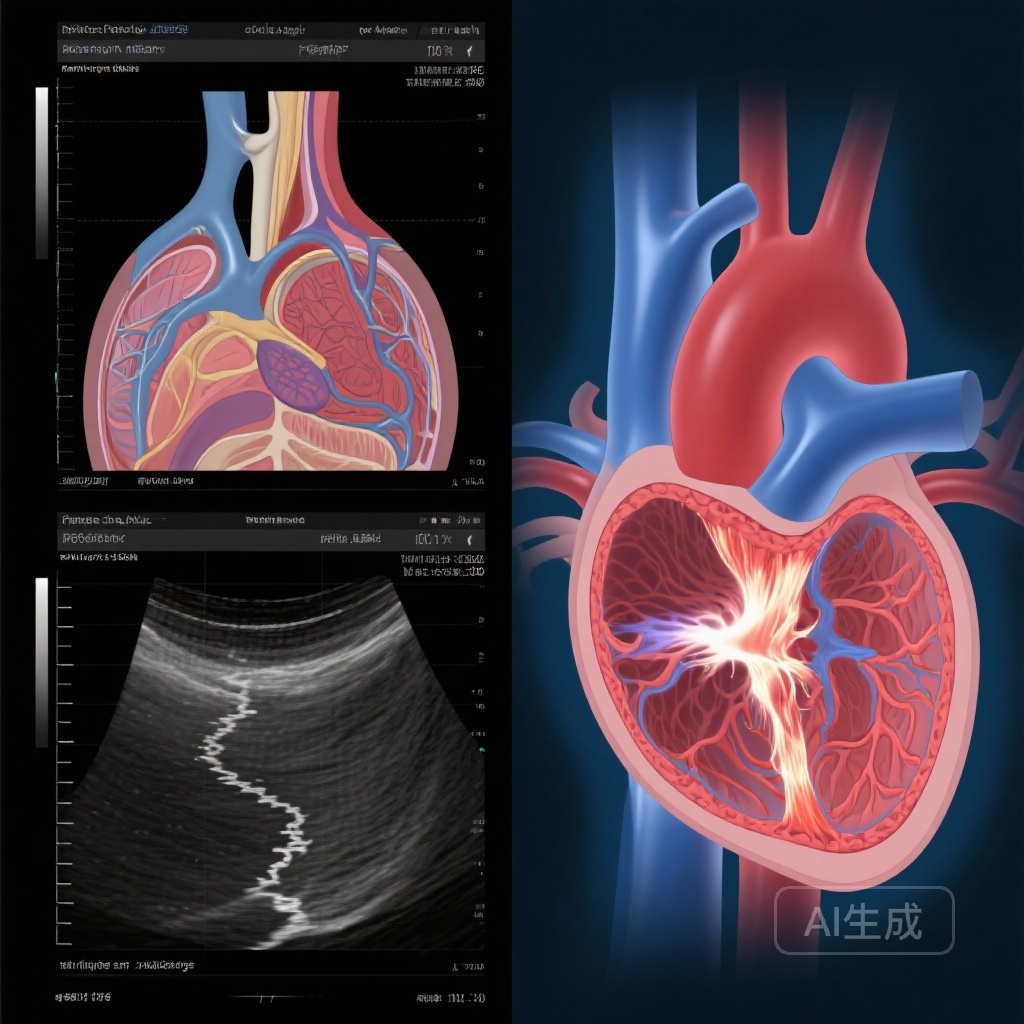
Chronic aortic regurgitation presents a persistent diagnostic challenge for cardiologists. While transthoracic echocardiography (TTE) remains the first-line imaging modality, quantifying AR severity accurately is fraught with technical limitations. The current gold standard for non-invasive quantitation is Cardiac Magnetic Resonance (CMR), which offers precise regurgitant volume (RegV) and fraction measurements. However, CMR is not always readily available, is costly, and cannot be performed at the bedside.
Within the realm of TTE, the Proximal Isovelocity Surface Area (PISA) method is the standard for quantifying valvular regurgitation. While highly effective for mitral regurgitation, its application in AR is often restricted by the difficulty of visualizing a hemispheric flow convergence zone from the apical window. This technical hurdle often leaves clinicians relying on qualitative or semi-quantitative measures, such as vena contracta width or pressure half-time, which can be influenced by loading conditions and ventricular compliance.
Study Design: A Head-to-Head Comparison with CMR
In a rigorous study recently published in the European Heart Journal – Cardiovascular Imaging, Zeitoun et al. sought to evaluate whether two-dimensional measurements of flow convergence (FC) from the standard parasternal long-axis (PLA) window could provide a more reliable assessment of AR severity. The study included 80 patients with chronic AR who underwent both TTE and CMR within a narrow four-hour window to ensure stable hemodynamic conditions.
The researchers focused on three specific FCPLA parameters:
1. Maximum Length (Lmax)
The maximum vertical extent of the flow convergence zone from the aortic valve plane into the left ventricular outflow tract.
2. Maximum Width (Dmax)
The maximum horizontal spread of the convergence zone.
3. Hemispheric Area
A calculated area based on the geometry of the flow convergence signal.
These parameters were compared against CMR-derived RegV, with significant AR defined as a RegV > 45 ml. The study also compared FCPLA performance against traditional markers, including vena contracta width (VCW), jet width/LVOT diameter ratio, descending aorta end-diastolic velocity, and pressure half-time (PHT).
Key Findings: Feasibility and Diagnostic Accuracy
The results revealed a striking difference in clinical utility between the parasternal and apical windows. While apical PISA quantitation was feasible in less than 40% of the cohort, FCPLA parameters were measurable in the vast majority (83%) of patients. This suggests that the parasternal window offers a superior acoustic path for visualizing the flow convergence zone in AR, likely due to the perpendicular orientation of the ultrasound beam to the regurgitant flow and the closer proximity of the valve to the transducer.
In terms of diagnostic accuracy, the FCPLA parameters showed impressive correlations with CMR RegV:
- Lmax: Spearman ρ = 0.75
- FCPLA Area: Spearman ρ = 0.78
- Dmax: Spearman ρ = 0.65
The statistical performance in identifying significant AR was particularly noteworthy. Using a threshold of Lmax = 0.55 cm, the AUC was 0.98. For an FCPLA area of 0.35 cm2, the AUC was 0.94. These values indicate a high degree of sensitivity and specificity, rivaling the accuracy of much more complex imaging protocols.
Comparative Performance Against Traditional Parameters
When compared to established echocardiographic markers, the FCPLA Lmax and Area parameters were found to be comparable to vena contracta width (AUC 0.94) and left ventricular end-diastolic volume index (AUC 0.91). Critically, they were statistically superior to several commonly used measures:
- Jet width/LVOT diameter (AUC 0.80)
- Descending aorta end-diastolic velocity (AUC 0.74)
- Pressure half-time (AUC 0.71)
This suggests that while PHT and jet width are useful screening tools, they lack the precision required for definitive grading of AR severity when compared to flow convergence in the PLA window.
Expert Commentary: Mechanistic Insights and Clinical Application
The success of the parasternal window in this study can be attributed to the physics of ultrasound. In the apical view, the flow convergence zone is often obscured by the high-velocity jet itself or shadowed by calcification of the aortic valve. In the parasternal long-axis view, the flow convergence occurs almost directly under the transducer, allowing for better resolution of the boundary between the laminar flow and the accelerating regurgitant stream.
From a clinical perspective, these findings provide a practical solution for the “indeterminate” AR cases often encountered in the echo lab. If the apical PISA is not visible—which is the case in 60% of patients according to this data—the clinician should immediately transition to the parasternal window to measure the Lmax of the flow convergence. An Lmax greater than 0.55 cm serves as a strong red flag for significant regurgitation that may warrant surgical consideration or closer CMR follow-up.
However, it is important to note the study’s limitations. As a single-center study with 80 patients, further multi-center validation is required before these specific cut-offs are integrated into formal societal guidelines. Furthermore, the presence of eccentric jets or multiple jets may complicate the 2D measurement of flow convergence, requiring a more nuanced approach in complex valvular pathologies.
Conclusion
The evaluation of aortic regurgitation is moving toward a more integrated approach, where bedside echocardiography is refined to match the accuracy of advanced cross-sectional imaging. This study demonstrates that flow convergence from the parasternal long-axis window is not only more feasible than apical PISA but also highly accurate in identifying significant AR. By incorporating FCPLA measurements—specifically Lmax and Area—into routine clinical practice, cardiologists can improve the reliability of AR grading, leading to more timely and appropriate interventions for patients with chronic valvular disease.
References
Zeitoun J, Attar R, Malahfji M, Angulo C, Shah DJ, Zoghbi WA. Evaluation of Aortic Regurgitation Severity Using Flow Convergence from the Parasternal Window: Comparison with Cardiac Magnetic Resonance and Traditional Doppler Parameters. Eur Heart J Cardiovasc Imaging. 2025 Dec 22:jeaf359. doi: 10.1093/ehjci/jeaf359. Epub ahead of print. PMID: 41430404.