Highlights
- Annexin A2 (ANXA2) is significantly upregulated in human and murine ischemic failing hearts, with circulating levels correlating positively with cardiac injury in patients.
- ANXA2 acts as a molecular brake on mitophagy by competitively inhibiting the interaction between the mitophagy receptor Prohibitin 2 (PHB2) and LC3B.
- The ANXA2-PHB2 interaction facilitates the proteasomal degradation of PHB2 via TRIM29-mediated K48-linked polyubiquitination.
- Inhibition or genetic depletion of cardiomyocyte-specific ANXA2 restores mitophagy, reduces oxidative stress, and significantly improves cardiac function and remodeling post-myocardial infarction.
The Burden of Ischemic Heart Disease and Mitochondrial Homeostasis
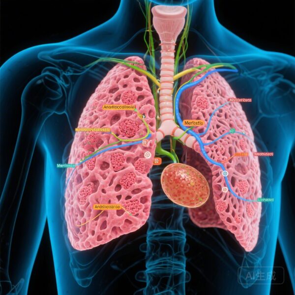
Myocardial infarction (MI) remains a leading cause of heart failure and mortality worldwide. Despite advances in reperfusion therapies, the pathological remodeling that follows acute ischemia often leads to progressive cardiac dysfunction. At the cellular level, the heart’s high metabolic demand makes it uniquely sensitive to mitochondrial health. Mitophagy—the selective autophagic degradation of damaged mitochondria—is a critical quality control mechanism that maintains cellular homeostasis during stress. However, the regulatory networks that govern mitophagy in the context of the infarcted heart are complex and, until recently, incompletely understood.
Recent research published in Circulation has identified Annexin A2 (ANXA2) as a pivotal negative regulator of cardiac mitophagy. While the Annexin family is known for its involvement in membrane trafficking and calcium signaling, this study elucidates a previously unknown role for ANXA2 in suppressing the PHB2-mediated mitophagy pathway, thereby exacerbating cardiac injury after MI.
Study Design and Methodology
To investigate the role of ANXA2, the research team employed a multi-faceted approach combining human clinical data, in vivo murine models, and in vitro cellular assays. Transcriptome analyses were first used to identify genes differentially expressed in the heart following ischemic injury. The clinical relevance was established by measuring circulating ANXA2 levels in patients with acute MI and comparing heart tissue from human ischemic failing hearts with non-failing controls.
The functional impact of ANXA2 was assessed using cardiomyocyte-specific Anxa2 knockdown and overexpression in mice. These models were subjected to permanent coronary artery ligation to simulate MI. Cardiac function was monitored via echocardiography, while infarct size and remodeling were evaluated through histological staining (Masson’s trichrome and Wheat Germ Agglutinin). In vitro experiments utilized neonatal rat ventricular myocytes (NRVMs) subjected to hypoxia to mimic ischemic conditions. Mechanistic insights were gained through immunoprecipitation (IP), mass spectrometry, and glutathione S-transferase (GST) pull-down assays to map protein-protein interactions.
Key Findings: ANXA2 as a Pathological Brake on Mitophagy
The study found that ANXA2 levels are dramatically elevated in both human and murine hearts following ischemic injury. Notably, in clinical cohorts, higher circulating ANXA2 levels were associated with more severe cardiac injury, suggesting its potential as a biomarker for disease progression. In animal models, the overexpression of ANXA2 in cardiomyocytes resulted in suppressed mitophagy, increased oxidative stress, and accelerated cell death, ultimately leading to worse cardiac remodeling and faster progression to heart failure.
Conversely, the depletion of ANXA2 proved to be highly cardioprotective. Mice lacking cardiomyocyte ANXA2 exhibited preserved mitochondrial function and enhanced mitophagy following MI. These animals showed significant reductions in infarct size, decreased inflammatory cell infiltration, and improved ejection fractions compared to wild-type controls. The protective effects of ANXA2 deficiency were directly linked to the maintenance of mitochondrial integrity and reduced reactive oxygen species (ROS) production.
Mechanistic Insight: The ANXA2-PHB2-TRIM29 Axis
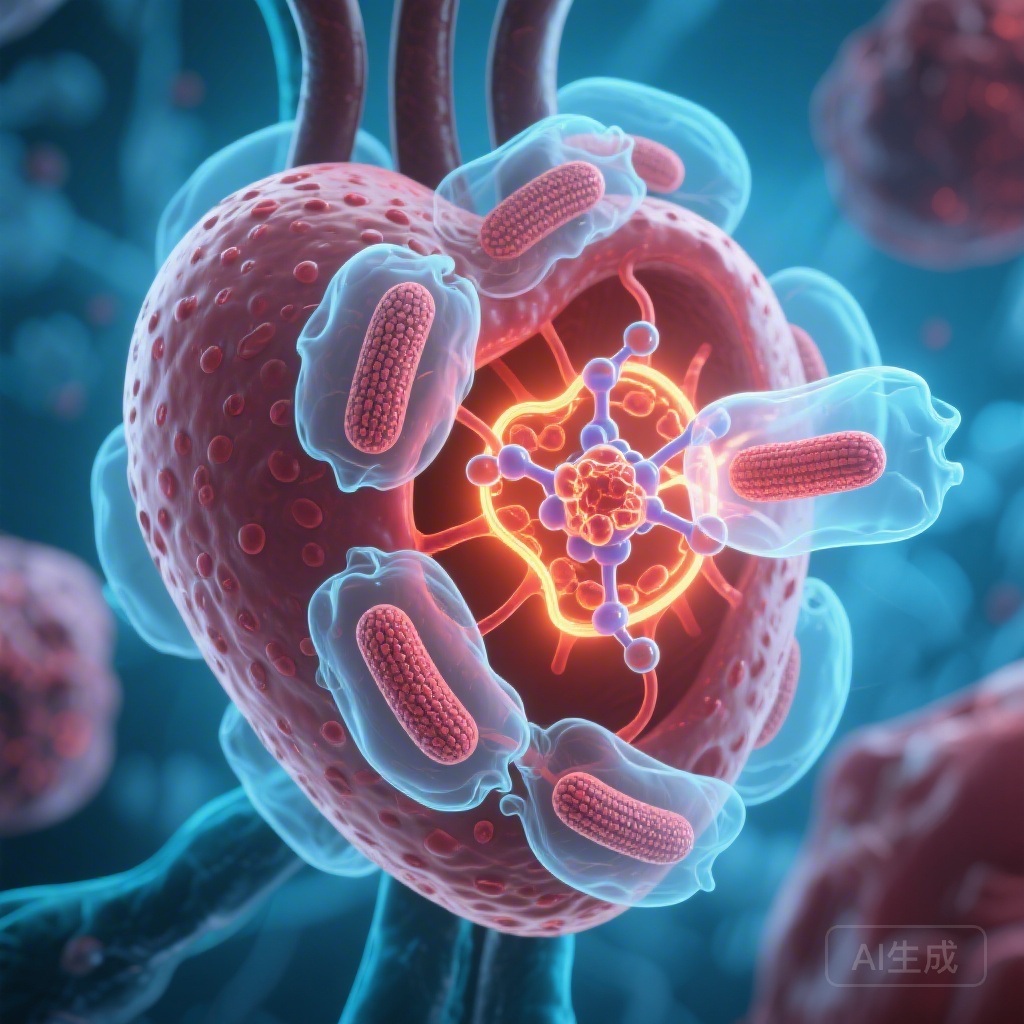
The most significant contribution of this research is the elucidation of the biochemical mechanism by which ANXA2 inhibits mitophagy. The researchers discovered that ANXA2 directly interacts with the mitophagy receptor Prohibitin 2 (PHB2) on the inner mitochondrial membrane. This interaction has two primary consequences:
1. Competitive Inhibition of LC3B Binding
Under normal mitophagy conditions, PHB2 acts as a bridge, binding to the autophagosomal protein LC3B to facilitate the engulfment of damaged mitochondria. ANXA2 competitively binds to PHB2, physically blocking the PHB2-LC3B interaction and effectively halting the mitophagy process.
2. Accelerated PHB2 Degradation
Furthermore, ANXA2 serves as a scaffold that recruits the E3 ubiquitin ligase TRIM29 (tripartite motif-containing 29) to PHB2. This recruitment leads to the K48-linked polyubiquitination of PHB2, marking it for proteasomal degradation. By reducing the total pool of available PHB2, ANXA2 ensures long-term suppression of mitochondrial clearance, leaving the cardiomyocyte vulnerable to the toxic effects of dysfunctional mitochondria.
The study confirmed this mechanism by showing that silencing Phb2 completely abolished the cardioprotective benefits of Anxa2 knockdown, proving that PHB2 is the essential downstream effector of ANXA2’s pathological action.
Expert Commentary and Translational Potential
The discovery of the ANXA2-PHB2-TRIM29 axis represents a significant leap in our understanding of cardiac proteostasis. For years, clinicians have sought ways to enhance mitophagy as a therapeutic strategy for heart failure. However, broad autophagy activators often lack specificity and can have off-target effects. Targeting ANXA2 offers a more nuanced approach by removing a specific “brake” that is upregulated only during pathological stress.
From a clinical perspective, the correlation between circulating ANXA2 and cardiac injury markers is particularly intriguing. If validated in larger cohorts, ANXA2 could serve as a predictive biomarker for post-MI remodeling, identifying patients at high risk of heart failure who might benefit from more aggressive intervention. Furthermore, small molecule inhibitors of the ANXA2-PHB2 interaction or inhibitors of TRIM29-mediated degradation could theoretically be developed to restore mitophagy in the acute phase of MI.
However, limitations remain. As ANXA2 is involved in other physiological processes, including fibrinolysis and membrane repair, systemic inhibition could carry risks. Future research must focus on cardiomyocyte-targeted delivery systems or specific disruptors of the ANXA2-PHB2 interface to minimize systemic toxicity.
Conclusion
The study by Deng et al. establishes ANXA2 as a critical checkpoint in cardiac mitochondrial quality control. By facilitating the degradation of the mitophagy receptor PHB2 and blocking its interaction with the autophagic machinery, ANXA2 prevents the clearance of damaged mitochondria, fueling the progression of heart failure after MI. The inhibition of ANXA2 represents a promising therapeutic strategy to restore mitochondrial homeostasis, alleviate cardiac injury, and improve the prognosis of patients suffering from ischemic heart disease.
References
Deng KQ, Xu Z, Wang Q, et al. Inhibition of Annexin A2 Facilitates PHB2-Mediated Mitophagy in Cardiomyocytes to Alleviate Cardiac Injury and Remodeling After Infarction. Circulation. 2026;153(11):826-844. PMID: 41492949.