Highlights
- Microvascular dropout (MvD) is a potent independent risk factor for structural progression in glaucoma suspects, with an odds ratio of 15.8.
- The progression of β-zone parapapillary atrophy (PPA) independently triples the risk of neuroretinal rim thinning or retinal nerve fiber layer (RNFL) defects.
- The combination of MvD and β-zone PPA progression yields a 75% predicted probability of structural optic disc damage.
- Structural changes often precede functional vision loss, occurring even in eyes that remain perimetrically stable for nearly two decades.
The Silent Progression of Glaucoma Suspects
Glaucoma management is often a race against time, where the goal is to intervene before irreversible vision loss occurs. However, for the millions of individuals classified as “glaucoma suspects,” the clinical path is often clouded by uncertainty. These patients may exhibit elevated intraocular pressure (IOP) or suspicious-looking optic discs, yet they maintain normal results on standard automated perimetry (SAP). The challenge for clinicians is identifying which of these suspects will experience structural decay and eventually progress to perimetric glaucoma.
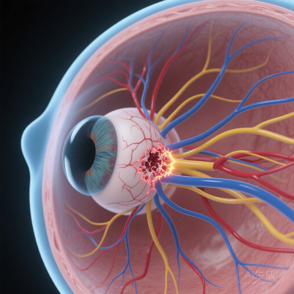
Recent advancements in imaging, specifically Optical Coherence Tomography Angiography (OCTA), have opened a new window into the vascular health of the optic nerve. One particular finding, microvascular dropout (MvD)—defined as a focal, complete loss of the deep-layer microvasculature in the parapapillary region—has emerged as a significant marker in established glaucoma. A recent study published in the American Journal of Ophthalmology by Soltani et al. investigates whether these microvascular and anatomical markers can predict structural progression in eyes that are still considered clinically stable.
Study Design and Methodology
In this retrospective cohort study, researchers analyzed 180 eyes from 134 glaucoma suspect patients. The rigor of the study is highlighted by its exceptional follow-up duration; fundus photographs were available for a mean period of 19.0 years (95% CI, 17.9 to 20.1). This long-term perspective is crucial for a disease as slowly progressive as glaucoma.
The study focused on three primary variables: structural progression, β-zone parapapillary atrophy (PPA) progression, and the presence of microvascular dropout (MvD). Structural progression was defined as new or enlarged RNFL defects or neuroretinal rim thinning, as graded by expert evaluation of stereophotographs. β-zone PPA progression was quantified by measuring changes in area, radial width, and angular extent, with a 20% increase in any parameter marking progression. Finally, MvD was assessed at the last visit using en-face choroidal vessel density maps derived from OCTA.
The inclusion criteria ensured that all eyes had at least five years of photographic history prior to the OCTA imaging, allowing researchers to relate late-stage microvascular status to nearly two decades of structural history.
Key Findings: Quantifying the Risk of Structural Decay
The results of the analysis provide a stark look at the risk factors driving optic disc damage. Of the 180 eyes studied, 58 (32.2%) were found to have MvD. The statistical associations were profound:
1. The Power of Microvascular Dropout
The presence of MvD was the strongest predictor of structural progression. Multivariable logistic regression showed that eyes with MvD had an odds ratio (OR) of 15.8 (95% CI, 5.6 to 44.6, P<0.001) for structural damage compared to those without. This suggests that the loss of the deep microvascular layer is not merely a late-stage byproduct of glaucoma but a marker closely tied to the ongoing destruction of the neuroretinal rim and RNFL.
2. Parapapillary Atrophy as a Mechanical Marker
β-zone PPA progression also proved to be a significant independent predictor, with an OR of 3.8 (95% CI, 1.2 to 11.7, P=0.022). PPA is often viewed as a sign of mechanical strain on the scleral tissue surrounding the optic nerve. Its expansion suggests that the structural support of the optic nerve head is being compromised, facilitating further axonal damage.
3. The Cumulative Effect
Perhaps the most clinically relevant finding was the additive effect of these markers. Margin plots estimated the predicted probability of structural progression based on these factors. Eyes with neither MvD nor PPA progression had a relatively low probability of structural change. However, for eyes exhibiting both MvD and β-zone PPA progression, the predicted probability of structural progression soared to 0.75. This high probability identifies a high-risk subgroup of suspects who may require more aggressive monitoring or earlier treatment.
Expert Commentary: Mechanistic Insights and Clinical Utility
The findings by Soltani et al. reinforce the dual-mechanism theory of glaucomatous damage, which posits that both vascular and mechanical factors contribute to optic nerve decay. The strong association of MvD suggests that perfusion deficits in the choriocapillaris or the deep parapapillary tissues may lead to chronic ischemia of the optic nerve head. When combined with the mechanical instability suggested by PPA progression, the optic nerve becomes highly vulnerable.
A critical takeaway for clinicians is that these structural changes were observed in eyes that remained “clinically stable” regarding their visual fields. This highlights the “pre-perimetric” window of opportunity. By the time a patient shows deficits on a visual field test, a significant percentage of retinal ganglion cells have already been lost. Identifying MvD through OCTA provides a non-invasive way to stratify risk before that functional loss occurs.
However, the study is not without limitations. As a retrospective analysis, it cannot definitively establish whether MvD precedes or follows structural thinning. Furthermore, MvD was only assessed at the final visit. Future longitudinal studies that track the emergence of MvD over time will be essential to determine if microvascular loss is a primary event or a secondary consequence of tissue atrophy.
Conclusion
The inclusion of OCTA into the clinical workflow for glaucoma suspects offers more than just pretty pictures; it provides actionable data. The presence of microvascular dropout, especially when paired with an expanding zone of parapapillary atrophy, serves as a major red flag for impending structural damage. For the physician-scientist and the clinician, these findings suggest that the definition of “stability” in glaucoma suspects needs to evolve. We must look beneath the surface at the microvascular integrity of the optic nerve to truly understand a patient’s risk profile and prevent the transition from suspect to sight-impaired.
References
1. Soltani G, Nishida T, Moghimi S, et al. Optic Disc Structural Progression in Glaucoma Suspect Eyes with Microvascular Dropout. American Journal of Ophthalmology. 2026. PMID: 41825843.
2. Weinreb RN, Aung T, Medeiros FA. The pathophysiology and treatment of glaucoma: a review. JAMA. 2014;311(18):1901-1911.
3. Richter GM, Madi I, Chu Z, et al. Structural and Vascular Predictors of Progression in Glaucoma Suspects and Early Glaucoma. Ophthalmology. 2020;127(10):1312-1322.