Introduction: The Challenge of Immunotherapy Resistance in HCC
Hepatocellular carcinoma (HCC) remains one of the most lethal malignancies globally, characterized by limited therapeutic options in its advanced stages. While immune checkpoint blockade (ICB), specifically targeting the PD-1/PD-L1 axis, has revolutionized the treatment landscape, the clinical reality remains sobering: a significant proportion of patients exhibit suboptimal responses or develop secondary resistance. The efficacy of ICB relies heavily on the presence and functionality of CD8+ T cells within the tumor microenvironment (TME). Recent research has highlighted a specific subset known as progenitor exhausted T (Tpex) cells. These cells serve as the ‘reservoir’ for the immune response; they possess stem-like properties, allowing them to self-renew and differentiate into effector-like cells upon ICB stimulation. However, in the harsh, immunosuppressive environment of HCC, these Tpex cells often rapidly progress toward a state of terminal exhaustion (Tex), where they lose their proliferative capacity and cytotoxic function. Understanding the molecular brakes that drive this transition is critical for improving patient outcomes.
Highlights
1. FYN was identified as a specific marker for progenitor exhausted T (Tpex) cells in HCC patients who respond successfully to immune checkpoint blockade.
2. Hyperactivation of the LCK kinase, particularly under high-affinity antigen stimulation, was found to drive Tpex cells toward terminal exhaustion by disrupting metabolic homeostasis.
3. LCK-mediated metabolic collapse is characterized by excessive glycolysis and impaired mitochondrial function, whereas FYN activity supports mitochondrial fitness and Tpex stemness.
4. Preemptive, low-dose LCK inhibition combined with anti-PD-1 therapy expands the Tpex cell pool and significantly improves therapeutic outcomes in preclinical models.
Uncovering the FYN/LCK Metabolic Switch
In a landmark study published in Hepatology, Wu et al. (2026) utilized an integrated analysis of single-cell RNA-sequencing (scRNA-seq) datasets to map the landscape of exhausted CD8+ T cells in HCC. Their objective was to identify the regulatory mechanisms that govern the maintenance of Tpex cells. The researchers discovered a critical divergence in the expression of two kinases belonging to the SRC family: FYN and LCK. While these kinases are closely related, they play distinct and often opposing roles in T-cell signaling and metabolism.
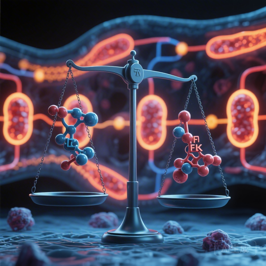
The study identified FYN as a hallmark of Tpex cells in patients who responded to ICB. Conversely, the absence or downregulation of FYN, coupled with the hyperactivation of LCK, was associated with terminal exhaustion. This led to the hypothesis that the FYN/LCK balance acts as a ‘metabolic switch’ that determines whether a T cell remains a stem-like progenitor or descends into irreversible exhaustion.
Study Methodology: From Single-Cell Atlases to Preclinical Validation
The researchers employed a multi-faceted approach to validate their findings. First, they constructed a comprehensive CD8+ T cell atlas using scRNA-seq data from HCC patients. This allowed them to track the differentiation trajectory of T cells from a naive state to progenitor exhaustion and, finally, to terminal exhaustion.
To explore the mechanistic role of these kinases, they utilized FYN-deficient and LCK-modulated CD8+ T cell models. High-affinity antigen stimulation was used to mimic the chronic TCR (T-cell receptor) signaling found in the tumor microenvironment. Metabolic profiling was conducted using Seahorse XF assays and mitochondrial imaging to assess glycolysis, oxidative phosphorylation (OXPHOS), and mitochondrial membrane potential. Finally, the team used preclinical HCC mouse models to test the therapeutic potential of modulating this axis. They explored two primary interventions: enhancing adoptive cell therapy (ACT) through transient LCK inhibition during T-cell expansion and preemptive low-dose LCK inhibition prior to administering anti-PD1 therapy.
Key Findings: How LCK Hyperactivation Drives Terminal Exhaustion
The most striking finding of the study was that LCK hyperactivation is a double-edged sword. While LCK is essential for initial T-cell activation, chronic and excessive LCK signaling—often triggered by high-affinity tumor antigens—accelerates terminal exhaustion. The researchers found that FYN deficiency directly induced this LCK hyperactivation.
When LCK becomes hyperactive, it triggers a cascade of metabolic changes that are detrimental to long-term T-cell survival. Specifically, it drives ‘metabolic overdrive,’ where the cell relies excessively on glycolysis. While glycolysis provides quick energy for effector functions, the lack of metabolic flexibility eventually leads to the accumulation of reactive oxygen species (ROS) and the collapse of mitochondrial function. These terminally exhausted cells showed fragmented mitochondria and a loss of mitochondrial DNA integrity, effectively ‘burning out’ their cellular engines.
In contrast, compensatory FYN activity was found to preserve mitochondrial fitness. FYN signaling appears to favor a more balanced metabolic profile, supporting oxidative phosphorylation and lipid metabolism, which are essential for the longevity and self-renewal of Tpex cells. By inhibiting LCK, the researchers were able to shift the balance back toward FYN, thereby restoring mitochondrial health and preventing the transition to terminal exhaustion.
Metabolic Reprogramming and Mitochondrial Fitness
The study provides deep mechanistic insights into how LCK disrupts the mitochondrial network. Hyperactive LCK signaling was linked to the dysregulation of key metabolic regulators, including the mTOR pathway and mitochondrial fusion/fission dynamics. In Tpex cells where the FYN/LCK balance was maintained, mitochondria remained elongated and functional, providing the energy required for sustained immune surveillance.
This ‘mitochondrial fitness’ is the cornerstone of Tpex stemness. Cells with robust mitochondria are better equipped to survive the nutrient-depleted and hypoxic conditions of the HCC tumor microenvironment. The study demonstrates that metabolic state is not just a byproduct of T-cell differentiation but a primary driver of it. By controlling the FYN/LCK switch, clinicians may be able to reprogram the metabolic ‘destiny’ of infiltrating T cells.
Translational Potential: LCK Inhibition as a Priming Strategy
The clinical implications of these findings are profound. The researchers tested the use of low-dose LCK inhibitors (such as dasatinib, which in low concentrations can selectively target LCK) in combination with standard immunotherapy.
In models of adoptive cell therapy, transiently inhibiting LCK during the ex vivo expansion phase resulted in a T-cell product with a much higher proportion of Tpex cells. When these cells were infused into HCC-bearing mice, they demonstrated superior persistence and anti-tumor efficacy compared to standard T cells.
Even more relevant for clinical practice was the ‘preemptive’ strategy. Administering low-dose LCK inhibition before anti-PD1 therapy effectively ‘primed’ the immune system. This treatment expanded the pool of Tpex cells within the tumor and prevented them from converting into terminal Tex cells once the anti-PD1 therapy was initiated. This sequential approach resulted in significantly smaller tumor volumes and prolonged survival in preclinical models, suggesting a way to overcome the primary resistance seen in many HCC patients.
Expert Commentary and Clinical Implications
This study represents a significant shift in how we view T-cell exhaustion in solid tumors. Traditionally, exhaustion was viewed as a linear and inevitable consequence of chronic antigen exposure. However, the identification of the FYN/LCK axis suggests that exhaustion is a regulated process that can be redirected through metabolic intervention.
From a clinical perspective, the use of LCK inhibitors requires careful calibration. LCK is fundamental to T-cell receptor signaling; complete or high-dose inhibition would result in immunosuppression. The ‘low-dose’ and ‘transient’ nature of the intervention described by Wu et al. is key. It acts more like a ‘tuner’ than an ‘off-switch,’ dampening excessive signals that lead to burnout while maintaining enough signaling for activation.
However, there are limitations to consider. The study primarily relies on preclinical models and retrospective scRNA-seq analysis. Transitioning these findings to human clinical trials will require precise dosing studies to ensure that the LCK inhibition does not interfere with the initial activation of naive T cells or the function of other immune subsets. Furthermore, the heterogeneity of HCC means that the FYN/LCK balance may vary significantly between patients, necessitating a biomarker-driven approach to patient selection.
Conclusion
The study by Wu et al. establishes the FYN/LCK kinase balance as a master regulator of T-cell fate in hepatocellular carcinoma. By acting as a metabolic switch, this axis governs the delicate transition between the regenerative Tpex state and the terminal Tex state. The finding that LCK inhibition can preserve mitochondrial fitness and stemness provides a novel therapeutic window to enhance the efficacy of both adoptive cell therapy and immune checkpoint blockade. As we move toward more personalized oncology, targeting the metabolic vulnerabilities of exhausted T cells may prove to be the key to unlocking long-term remissions in HCC.
References
1. Wu J, Jiang Z, Li Q, et al. FYN/LCK kinase balance as a metabolic switch orchestrates progenitor exhausted T Cell differentiation in hepatocellular carcinoma. Hepatology. 2026; doi: 10.1097/HEP.0000000000000XXX. (PMID: 41812035).
2. Miller BC, Sen DR, Al Abosy R, et al. Subsets of exhausted CD8+ T cells differentially mediate tumor control and respond to checkpoint blockade. Nature Immunology. 2019;20(3):326-336.
3. Im SJ, Hashimoto M, Gerner MY, et al. Defining CD8+ T cells that provide the proliferative burst after PD-1 therapy. Nature. 2016;537(7620):417-421.
4. Finn RS, Qin S, Ikeda M, et al. Atezolizumab plus Bevacizumab in Unresectable Hepatocellular Carcinoma. New England Journal of Medicine. 2020;382(20):1894-1905.