Highlights
Research published in the European Heart Journal (2026) has identified PlexinD1 as a critical mechanosensitive regulator of macrophage behavior in atherosclerosis. Key findings include:
1. Myeloid-specific PlexinD1 deficiency significantly attenuates the progression of atherosclerotic lesions, particularly at sites characterized by disturbed flow and oscillatory shear stress (OSS).
2. Oscillatory shear stress promotes classical M1 macrophage polarization by down-regulating the endothelial PTGS2/PGE2 pathway, which in turn releases the inhibition on PlexinD1/NF-κB signaling.
3. Clinical data from patients with acute coronary syndrome (ACS) show a strong correlation between elevated plasma PlexinD1 levels and the presence of high-risk coronary bifurcation lesions.
4. Novel PlexinD1-targeted multi-modal nanoparticles provide a viable platform for the non-invasive identification and monitoring of vulnerable plaques in vivo.
Background: The Hemodynamic Challenge in Atherosclerosis
Atherosclerosis is not a stochastic disease; it exhibits a striking spatial preference for specific anatomical sites. While linear segments of arteries exposed to laminar shear stress (LSS) remain relatively protected, arterial bifurcations, curvatures, and branch points are highly susceptible. These areas are characterized by disturbed flow and oscillatory shear stress (OSS), which promote endothelial dysfunction and chronic vascular inflammation. Despite the well-known impact of hemodynamics on endothelial cells, the mechanisms by which these physical forces are translated into macrophage-driven inflammation—the hallmark of plaque vulnerability—have remained poorly understood. Current lipid-lowering therapies, such as statins and PCSK9 inhibitors, significantly reduce systemic risk but often fail to address the localized inflammatory cascades triggered by local mechanical environments. This unmet clinical need has driven the search for ‘mechanoinflammatory’ targets that could specifically stabilize plaques at high-risk bifurcation sites.
Study Design and Methodology
To investigate the role of PlexinD1 in this context, researchers employed a comprehensive multi-pronged approach combining clinical observation, genetically modified animal models, and advanced molecular imaging. The clinical component involved a study of 72 patients diagnosed with acute coronary syndrome (ACS). These patients were stratified based on whether their lesions involved coronary bifurcations, and their plasma PlexinD1 levels were quantified and compared. Additionally, human tissue samples from carotid bifurcations (OSS zones) and proximal common carotid arteries (LSS zones) were analyzed for PlexinD1 expression and macrophage markers.
In the laboratory, the team generated myeloid-specific PlexinD1 knockout mice (PlexinD1-MKO) on an apolipoprotein E-deficient (ApoE-/-) background. These mice were subjected to a high-fat, high-cholesterol diet to induce atherosclerosis. To simulate the different flow conditions, researchers used co-culture systems of endothelial cells and macrophages, subjecting them to controlled LSS or OSS in vitro. Finally, to explore diagnostic potential, the study developed PlexinD1-targeted multi-modal nanoparticles designed for both optical and magnetic resonance imaging, testing their ability to detect vulnerable lesions in the mouse models.
Key Findings: PlexinD1 as a Mechanistic Switch
Clinical Correlations in Human Patients
The clinical data revealed that patients with coronary bifurcation lesions exhibited a 1.32-fold higher level of plasma PlexinD1 compared to those with non-bifurcation lesions. This increase was mirrored in histological analyses of human carotid plaques. Plaques located at the carotid bifurcation—exposed to OSS—demonstrated significantly higher levels of PlexinD1 expression and M1 macrophage polarization (marked by CD80 and iNOS) compared to plaques in the common carotid artery. These OSS-exposed plaques also showed features of higher vulnerability, such as larger necrotic cores and thinner fibrous caps.
Myeloid-PlexinD1 Deficiency and Plaque Attenuation
The animal studies provided definitive evidence of PlexinD1’s causal role. In ApoE-/- mice, the deletion of PlexinD1 specifically in the myeloid lineage led to a marked reduction in atherosclerotic lesion area. More importantly, the plaques that did form in the knockout mice were more stable, characterized by reduced M1 macrophage infiltration and increased markers of M2 (anti-inflammatory) polarization. This suggests that PlexinD1 is essential for the transition of macrophages into a pro-inflammatory state under the influence of disturbed flow.
The PTGS2/PGE2/PlexinD1/NF-κB Axis
Mechanistically, the study uncovered a sophisticated cross-talk between endothelial cells and macrophages. Under healthy laminar flow (LSS), endothelial cells produce high levels of Prostaglandin-Endoperoxide Synthase 2 (PTGS2) and Prostaglandin E2 (PGE2). This PGE2 acts on macrophages to suppress PlexinD1 expression. However, when exposed to oscillatory shear stress (OSS), endothelial PTGS2/PGE2 production is down-regulated. This reduction in PGE2 allows for the up-regulation of PlexinD1 in macrophages, which subsequently activates the NF-κB signaling pathway. This cascade ultimately drives the classical M1 polarization that fuels plaque progression and destabilization.
Diagnostic Innovation: Targeted Nanoparticles
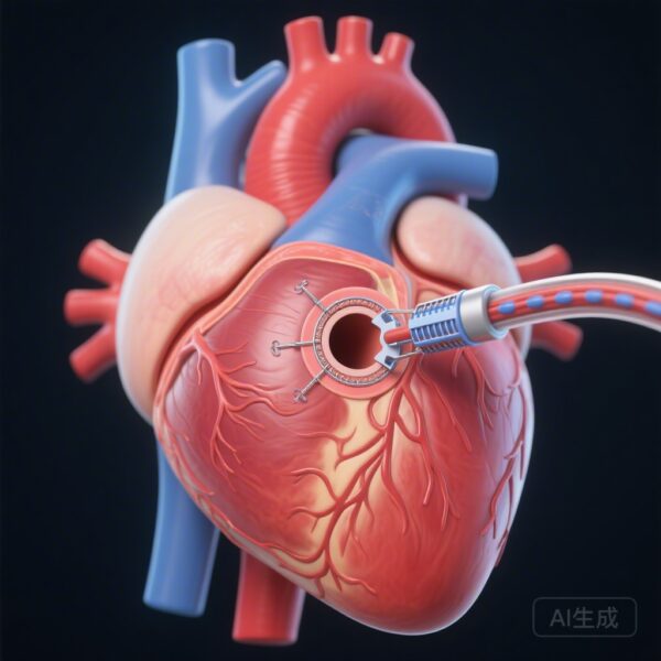
The development of PlexinD1-targeted multi-modal nanoparticles represented a significant translational achievement. These nanoparticles were able to selectively accumulate in bifurcation lesions in the mouse models, providing clear signals for both fluorescence and MRI. This demonstrates that PlexinD1 is not only a therapeutic target but also a highly specific biomarker for identifying ‘hot’ plaques that are at high risk of rupture.
Expert Commentary and Clinical Implications
The discovery of the PlexinD1-mediated mechanoinflammatory pathway represents a significant shift in our understanding of atherosclerosis. Traditionally, macrophages were viewed as passive responders to lipid accumulation. This study positions them as active mechanosensors that integrate signals from the overlying endothelium to dictate the inflammatory tone of the vessel wall. From a clinical perspective, this provides a potential explanation for why some plaques remain stable while others, often at bifurcations, progress rapidly toward rupture.
While the results are promising, certain limitations remain. The use of mouse models, while standard, does not perfectly replicate the complex, multi-decade progression of human atherosclerosis. Furthermore, the systemic effects of targeting PlexinD1 must be carefully evaluated, as Plexin signaling is involved in various physiological processes, including neurodevelopment and angiogenesis. However, the use of targeted nanoparticles suggests a path toward localized therapy or high-precision diagnostics that could minimize systemic side effects. Future research should focus on whether PlexinD1 levels can predict clinical outcomes in larger prospective cohorts and whether pharmacological inhibition of this pathway can stabilize existing high-risk lesions in humans.
Conclusion
In summary, the study by Ma et al. identifies PlexinD1 as a pivotal mediator of atherosclerosis at sites of disturbed flow. By suppressing the endothelial PTGS2/PGE2 brake, oscillatory shear stress activates a PlexinD1/NF-κB-dependent inflammatory program in macrophages. Myeloid-PlexinD1 deficiency effectively halts this process, leading to smaller and more stable atherosclerotic plaques. As we move toward an era of precision cardiovascular medicine, PlexinD1 emerges as a dual-purpose target for both the sophisticated imaging of vulnerable plaques and the development of novel immunomodulatory therapies.
References
Ma M, Zhang Y, Gao L, Wang Z, Xin R, Wang M, Zhang C, Sun Z, Liu L, Hui H, Tian J, Chen Y. Atherosclerotic progression at sites of low shear stress is attenuated by myeloid-PlexinD1 deficiency through suppression of classical macrophage polarization. European Heart Journal. 2026-Mar-09;47(10):1242-1259. PMID: 41405851.