Introduction: The Changing Landscape of Assisted Reproductive Technology
The utilization of Assisted Reproductive Technology (ART) has seen a paradigm shift over the last decade, particularly with the rise of the freeze-all strategy. As frozen embryo transfer (FET) becomes increasingly common due to improved cryopreservation techniques like vitrification and the potential for better endometrial synchrony, clinical focus has expanded beyond success rates to the long-term health of the offspring. It is well-documented that FET is associated with higher birthweights compared to fresh embryo transfer (fresh-ET) and natural conception (NC). However, the downstream physiological consequences of these altered early growth trajectories—specifically regarding skeletal development and metabolic health—remain areas of intense investigation. Two recent landmark studies provide a comprehensive look at these outcomes, evaluating bone mineralization and body mass index (BMI) in children conceived through various ART methods.
Highlighting Key Insights
1. Children conceived after FET demonstrate a significantly higher height-adjusted bone mineral content (BMC) at age 7-10 compared to those from fresh-ET or natural conception.
2. The observed increase in bone mineralization in FET offspring is primarily mediated by their higher birthweight, rather than inherent metabolic differences in bone turnover markers like calcium or parathyroid hormone (PTH).
3. Systematic review and meta-analysis of over 32 studies indicate no clinically significant difference in BMI standard deviation scores (SDS) between ART-conceived children and naturally conceived peers from ages 1 to 18.
4. While FET is linked to a higher risk of being born large-for-gestational-age (LGA), the long-term metabolic profile regarding BMI appears reassuringly stable across childhood and adolescence.
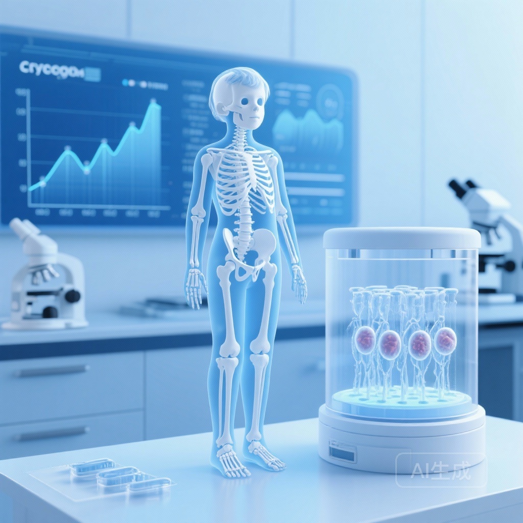
Bone Mineralization in the HiCART Cohort
Study Design and Methodology
The ‘Health in Childhood following Assisted Reproductive Technology’ (HiCART) study was a retrospective cohort study conducted in Denmark. It analyzed 606 singletons, including 200 conceived via FET, 203 via fresh-ET, and 203 via natural conception. The children, born between 2009 and 2013, underwent clinical examination at 7-10 years of age. The researchers utilized whole-body dual-energy x-ray absorptiometry (DXA) scans—the gold standard for assessing bone health—to measure bone mineral content (BMC). Importantly, the analysis adjusted for height, as BMC is highly dependent on body size. Secondary measures included fasting blood samples to assess bone-regulating hormones and growth factors, such as IGF-1, calcium, and PTH.
Key Findings: The Role of Birthweight
The crude data initially suggested no major differences in BMC across the groups. However, after adjusting for relevant confounders (such as maternal age, BMI, and smoking), the FET group showed a statistically significant increase in height-corrected BMC compared to both the fresh-ET and NC groups.
The most striking finding emerged when birthweight (BW) was introduced into the model. Once adjusted for birthweight SD scores, the statistical significance of the increased BMC in FET children vanished. This suggests that the skeletal benefits—or differences—seen in middle childhood are not an independent effect of the freezing process itself on bone metabolism, but rather a consequence of the increased fetal growth and higher birthweight commonly observed in FET pregnancies. Interestingly, there were no differences in lean mass, physical activity levels, or biochemical markers of bone metabolism (calcium, PTH, IGF-1) between the three groups, reinforcing the theory that the difference is structural and growth-related.
Longitudinal BMI Trends: A Systematic Meta-Analysis
Evaluating the Risk of Childhood Obesity
Parallel to skeletal health, there has been ongoing concern that the epigenetic modifications potentially induced by ART could influence metabolic programming, leading to increased risks of obesity. A comprehensive systematic review and meta-analysis (Asserhøj et al., 2025) addressed this by examining BMI in children aged 1-18 years. The review included 32 studies meeting strict inclusion criteria after screening over 22,000 records.
Meta-Analysis Results
The researchers conducted three primary meta-analyses:
1. ART vs. Natural Conception (BMI SDS): Analysis of 8,902 ART children and 61,818 NC children showed no difference in BMI SDS (mean difference 0.02; 95% CI: -0.03 to 0.06). This is a highly reassuring finding for parents and clinicians alike.
2. ART vs. Natural Conception (Raw BMI): A second analysis of 41,530 children showed a very slight decline in raw BMI (-0.16 kg/m2) in ART children compared to NC, which, while statistically significant, is likely not clinically relevant.
3. FET vs. Fresh-ET (BMI SDS): Comparing the two main ART modalities (n=20,855), no significant difference in BMI SDS was found (mean difference 0.08; 95% CI: -0.02 to 0.18).
Despite the heterogeneity of the studies (I2 values ranging from 9% to 87%), the overall trend suggests that the birthweight variations associated with FET (higher weight) and fresh-ET (lower weight) do not necessarily translate into a higher BMI in later childhood. This mitigates some concerns that FET-induced LGA or fresh-ET-induced SGA (small-for-gestational-age) would lead to a significant surge in childhood obesity through rapid catch-up growth or metabolic over-compensation.
Expert Commentary: Mechanistic Insights and Clinical Implications
The finding that FET children have higher BMC mediated by birthweight is a critical piece of the developmental programming puzzle. In natural conception, birthweight is a known predictor of adult bone mass. The fact that FET children maintain this relationship suggests that the “extra” growth achieved in utero during FET pregnancies translates into a sturdier skeletal frame by age 10.
However, several questions remain. Why does FET lead to higher birthweight? Hypotheses include the influence of the cryoprotectants used, the process of freezing and thawing itself, or the different hormonal environment of a prepared endometrium in FET cycles compared to the hyper-stimulated environment of fresh-ET cycles. The lack of difference in IGF-1 and PTH levels suggests that the skeletal differences are established early in development and maintained through childhood growth rather than being driven by ongoing hormonal alterations.
Regarding BMI, the results are largely positive. While previous studies have occasionally suggested a higher risk of metabolic syndrome in ART offspring, this large-scale meta-analysis provides moderate-certainty evidence that BMI—a primary marker for metabolic health—is not significantly adversely affected. However, the authors of the review correctly point out that BMI is a surrogate measure; it does not distinguish between fat mass and lean mass. Future research should prioritize more granular body composition analysis, such as the DXA-based lean and fat mass measurements used in the HiCART study.
Limitations and Future Directions
Both studies acknowledge limitations that warrant caution. In the bone mineralization study, the specific cause of parental infertility was not available, leaving room for residual confounding. Additionally, physical activity was assessed via parental questionnaires, which are prone to information bias. In the BMI meta-analysis, many included studies were classified as low or very low quality, and high heterogeneity was observed in comparisons between FET and fresh-ET.
Longitudinal studies that follow FET and fresh-ET cohorts into late adolescence and early adulthood are essential. We need to determine if the increased BMC in FET children persists after puberty and if it eventually results in a higher peak bone mass, which would be protective against osteoporosis later in life. Similarly, while BMI appears stable, the risk of cardiovascular dysfunction or glucose intolerance in the ART population requires ongoing vigilance.
Conclusion
For clinicians and patients navigating the complexities of ART, these findings are predominantly encouraging. The increased bone mineral content in FET-conceived children appears to be a robust, growth-mediated phenomenon that follows the trajectory established by their higher birthweights. Furthermore, the stability of BMI across the ART population suggests that the concerns regarding a surge in childhood obesity related to embryo freezing may be overstated. As the first generation of FET-conceived children reaches adulthood, continued monitoring will be vital to confirming these positive health indicators across the lifespan.
Funding and ClinicalTrials.gov
The HiCART study was supported by the Novo Nordisk Foundation (grant numbers: NNF18OC0034092, NFF19OC0054340) and the Rigshospitalet Research Foundation. The study is registered at ClinicalTrials.gov under the identifier NCT03719703. The systematic review on BMI was registered with PROSPERO (CRD42021257788).
References
1. Grønlund EW, Kjaer ASL, Asserhøj LL, et al. Bone mineralization in children aged 7-10 years born after ART with frozen and fresh embryo transfer. Hum Reprod Open. 2025;2025(4):hoaf055. doi:10.1093/hropen/hoaf055.
2. Asserhøj LL, Kielstrup LR, Grønholdt CL, et al. BMI in children aged 1-18 years conceived after ART with fresh and frozen embryo transfer: a systematic review and meta-analyses. Hum Reprod Update. 2025;31(6):643-671. doi:10.1093/humupd/dmaf017.