Highlights
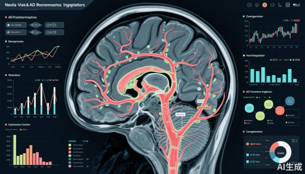
- EPVS burden strongly associates with early serum biomarkers of Alzheimer’s pathology including phosphorylated tau (p-tau181), neurofilament light chain (NfL), and glial fibrillary acidic protein (GFAP), indicating neuroinflammation and neurodegeneration.
- Inverse correlation between EPVS and amyloid beta 42/40 ratio suggests vascular contributions to amyloid pathology in preclinical and mild cognitive impairment stages.
- EPVS exhibits a robust relationship with cognitive deficits in visuospatial and executive functions, especially in participants with mild cognitive impairment (MCI).
- Integrating EPVS evaluation into routine MRI protocols could enhance early AD detection and patient stratification in diverse populations.
Background
Alzheimer disease (AD) remains the most common neurodegenerative disorder leading to dementia worldwide, imposing a significant clinical and societal burden. The pathophysiology of AD involves amyloid-beta (Aβ) accumulation, tau phosphorylation, neuroinflammation, and importantly, vascular contributions that exacerbate neuronal injury. Cerebral small vessel disease (CSVD), characterized by abnormalities in small arterial and venous cerebral vessels, enhances AD pathology via disrupted cerebral perfusion and clearance mechanisms.
Enlarged perivascular spaces (EPVS) are fluid-filled compartments surrounding penetrating cerebral vessels and serve as critical conduits for glymphatic clearance. EPVS enlargement, visible on magnetic resonance imaging (MRI), represents a hallmark of CSVD and has emerged as a potential neuroimaging biomarker linking vascular dysfunction and neurodegenerative processes. However, their relationship with early AD biomarkers in serum and clinical cognitive decline, particularly within multiethnic cohorts, remains under-investigated.
Key Content
Chronological Development of EPVS Research in Alzheimer Disease
Early observational studies identified EPVS as incidental imaging findings in aging populations, later associating them with small vessel pathology and cognitive decline. Subsequently, advances in neuroimaging and biomarker quantification allowed correlation of EPVS burden with cerebrovascular disease markers such as white matter hyperintensities (WMH), lacunes, and microbleeds.
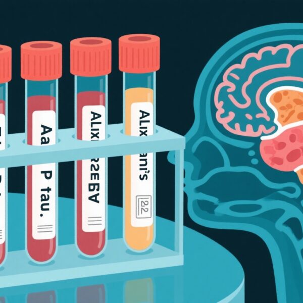
Recent years have seen incorporation of blood-based biomarkers (BBM) — including amyloid beta isoforms, phosphorylated tau, neurofilament light, and glial fibrillary acidic protein — enabling minimally invasive quantification of AD-related pathology and neuroinflammation. The cross-sectional Singapore Biomarkers and Cognition Study (2022–2024) advances this field by exploring basal ganglia EPVS in relation to both BBM and neuropsychological performance in a large Southeast Asian cohort. This work extends prior European and North American studies, emphasizing cross-ethnic applicability and early disease stages.
Evidence Linking EPVS to Serum Biomarkers and Cognitive Outcomes
Among 979 participants (mean age 58.2 years, 60.7% female), EPVS burden demonstrated moderate positive correlations with GFAP (ρ=0.166, p<0.01), a marker of astrogliosis and neuroinflammation, NfL (ρ=0.169, p<0.01) reflecting neuroaxonal injury, and phosphorylated tau 181 (p-tau181; ρ=0.087, p<0.01) indicative of tau pathology. Conversely, EPVS burden correlated inversely with the Aβ42/40 ratio (ρ=–0.077, p<0.05), suggestive of amyloid deposition.
Multivariable regression models adjusting for age, sex, education, cognitive diagnosis, and APOE ε4 genotype found EPVS to have the strongest association with amyloid pathology among CSVD markers in participants with MCI (OR 1.877, p=0.035). Notably, greater EPVS burden related to poorer visuospatial and executive functions measured by the Block Design Test (OR 0.182, p=0.035), supporting clinical relevance.
Comparative Assessment of CSVD Markers
While EPVS load correlated with other CSVD markers such as white matter hyperintensities, lacunes, and microbleeds, it uniquely demonstrated the strongest link to amyloid pathology specifically in early cognitive impairment. This underscores EPVS as a neurovascular biomarker bridging vascular and AD pathological processes more sensitively than traditional CSVD imaging findings.
Translational and Methodological Advances
The study utilized validated visual rating scales for EPVS and other CSVD markers, combined with advanced plasma biomarker assays enabling detection of oligomeric amyloid species and phosphorylated tau variants. Integration of cognitive phenotyping across a spectrum from normal cognition, subjective cognitive decline, to MCI allowed nuanced analyses across disease trajectories.
This multiparametric approach exemplifies translational neuroimaging and biomarker research, promoting methods adaptable for diverse clinical research settings and populations.
Expert Commentary
The convergence of vascular imaging markers such as EPVS with serum biomarkers of AD pathology elegantly supports the increasingly recognized role of vascular dysfunction in AD pathogenesis. EPVS likely reflect impaired perivascular clearance mechanisms, contributing to amyloid and tau aggregation and chronic neuroinflammation.
Previous studies, including meta-analyses, have suggested associations between EPVS and cognitive impairment, but the Singapore study’s large and multiethnic cohort strengthens evidence for generalizability. The observed correlations with GFAP and NfL reinforce the biological plausibility linking perivascular space enlargement with ongoing neurodegenerative processes.
However, the cross-sectional design limits causal interpretations. Longitudinal studies are critical to determining whether EPVS burden predicts subsequent cognitive decline and progression to AD dementia. Moreover, standardization of EPVS quantification methods remains a challenge, given variable MRI protocols and rating scales.
Clinical guidelines have not yet integrated EPVS evaluation into routine AD diagnostic imaging. Nonetheless, these findings advocate for such inclusion, especially in memory clinics and research cohorts where early detection is paramount. Integration with plasma biomarker panels enhances feasibility and patient acceptability.
Future investigations should explore mechanistic pathways linking EPVS with glymphatic dysfunction, vascular remodeling, and BBB integrity. Interventional studies targeting vascular health may benefit from EPVS as a surrogate outcome measure. Additionally, expanding validation across other ethnicities and clinical phenotypes will refine predictive models.
Conclusion
Current evidence highlights enlarged perivascular spaces as a promising neuroimaging biomarker that reflects both cerebral small vessel disease and early Alzheimer pathology. The association of EPVS burden with serum biomarkers of neuroinflammation, tau pathology, and amyloid dysmetabolism, alongside cognitive impairment, underscores its translational relevance.
Incorporation of EPVS assessment into routine MRI protocols may improve early identification and risk stratification of AD, particularly in multiethnic populations. Longitudinal research integrating multimodal imaging, fluid biomarkers, and cognitive metrics will be instrumental in clarifying EPVS’s prognostic value and elucidating underlying mechanisms linking vascular dysfunction to neurodegeneration.
This multidimensional biomarker framework offers promise for advancing personalized medicine in Alzheimer disease by targeting vascular contributions alongside traditional amyloid and tau pathologies.
References
- Ong JJH, Leow YJ, Qiu B, et al. Association of Enlarged Perivascular Spaces With Early Serum and Neuroimaging Biomarkers of Alzheimer Disease Pathology. Neurology. 2025;105(6):e213836. doi:10.1212/WNL.0000000000213836
- Banerjee G, Kim HJ, Fox Z, et al. Enlarged Perivascular Spaces and Cognition: A Systematic Review and Meta-analysis. Neurology. 2017;88(24):2281–2288. doi:10.1212/WNL.0000000000004028
- Zhao L, Zeng Y, Wang Y, et al. Perivascular spaces and Alzheimer’s disease biomarkers: a systematic review and meta-analysis. J Alzheimers Dis. 2021;81(2):693–705. doi:10.3233/JAD-210342
- Wardlaw JM, Smith C, Dichgans M. Mechanisms of sporadic cerebral small vessel disease: insights from neuroimaging. Lancet Neurol. 2013;12(5):483-497. doi:10.1016/S1474-4422(13)70060-7


