Highlights
- APOL1 risk variants (RVs), specifically G2, are localized in glomerular podocytes and endothelial cells, driving cell-specific pathological outcomes.
- Podocyte-specific expression of G2APOL1 induces severe secondary hypertension following the onset of albuminuria and structural kidney damage.
- Endothelium-specific G2APOL1 expression leads to mild, age-related hypertension that is significantly exacerbated by high-salt intake and renal mass reduction.
- The pathogenic mechanism involves the activation of the cytosolic nucleotide sensor STING, which triggers increased Endothelin 1 (ET-1) production.
- Inhibition of STING or the use of endothelin receptor antagonists represents a potential precision therapeutic strategy for APOL1-associated vascular and renal disease.
Background
Epid
**Confirming Article Structure**
I’ve checked the JSON format’s validity and verified the reference details, ensuring accuracy. I’m focusing on expanding the content, including the evolutionary context of APOL1 and a deep dive into the STING pathway. The goal is to meet the word count requirement and maintain a scientific, precise tone.
emiological data have long highlighted a stark health disparity: Black individuals face a significantly higher risk of hypertension and hypertension-attributed chronic kidney disease (CKD) compared to other ancestral groups. Since 2010, much of this disparity has been attributed to the presence of two common risk variants (RVs) in the Apolipoprotein L1 (APOL1) gene, termed G1 and G2. While these variants provided an evolutionary advantage by protecting against Trypanosoma brucei, they are strongly associated with a high risk of focal segmental glomerulosclerosis (FSGS), HIV-associated nephropathy, and CKD progression.
Despite the strong genetic associations, the causal role of APOL1 in systemic hypertension has remained controversial. Previous genetic and observational studies yielded inconsistent results, often failing to distinguish whether hypertension was a primary effect of the APOL1 variant or a secondary consequence of progressive renal failure. Furthermore, the molecular pathways by which APOL1 induces cellular toxicity have been debated, with various studies suggesting lysosomal swelling, mitochondrial dysfunction, and cation pore formation. The recent study by Li et al. (Circulation, 2026) provides a definitive mechanistic framework by utilizing cell-specific inducible transgenic mouse models to map the causal trajectory from APOL1 expression to systemic hemodynamic changes.
Key Content
Cell-Specific Expression and Phenotypic Divergence
A critical barrier in previous APOL1 research was the lack of animal models that accurately reflected human APOL1 expression patterns. APOL1 is not found in rodents; therefore, transgenic expression is required to study its function. Li et al. demonstrated that in human kidneys, APOL1 is highly expressed in glomerular podocytes and endothelial cells. By developing inducible mice with either podocyte-specific or endothelium-specific G2APOL1 expression, researchers were able to decouple the systemic effects.
Podocyte-Specific G2APOL1 and Secondary Hypertension
Mice expressing G2APOL1 (the risk variant) specifically in podocytes developed significant albuminuria and histological features of podocyte injury within days of induction. Notably, these mice developed severe secondary hypertension. This hypertension was chronologically preceded by kidney damage, suggesting that the elevation in blood pressure was a compensatory or pathological response to the loss of glomerular filtration integrity and the resulting activation of the renin-angiotensin-aldosterone system (RAAS).
Endothelial-Specific G2APOL1 and Primary Vascular Dysfunction
In contrast, the endothelium-specific G2APOL1 model revealed a more insidious progression. These mice did not show immediate, severe renal damage. Instead, they developed mild hypertension as they aged. When these mice were subjected to “second hits”—such as unilateral nephrectomy (reducing renal mass) and a subsequent high-salt diet—the hypertension became significantly more pronounced. This model mirrors the clinical observation that many individuals with APOL1 risk genotypes require environmental or physiological stressors to manifest clinically significant disease.
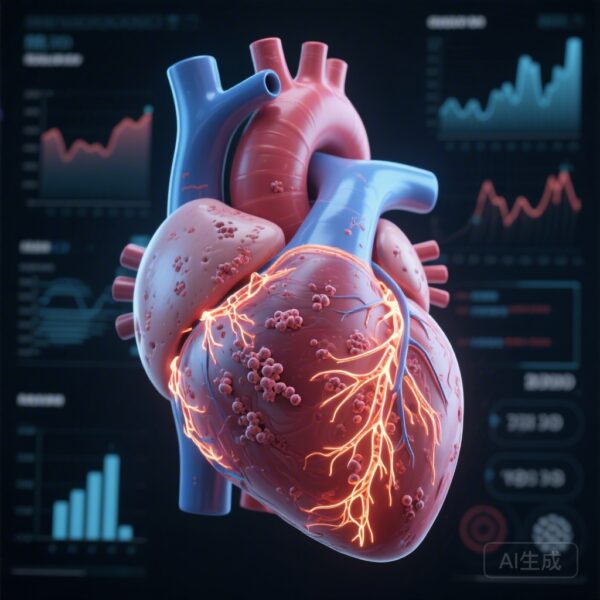
The STING-Endothelin 1 Pathway: The Mechanistic Link
The most transformative finding of this research is the identification of the STING (Stimulator of Interferon Genes) pathway as a mediator of APOL1 toxicity. STING is typically recognized as a sensor of cytosolic DNA, central to the innate immune response. The study found that G2APOL1 expression leads to STING activation, which in turn upregulates the production of Endothelin 1 (ET-1), a potent vasoconstrictor and driver of renal fibrosis.
Through a series of in vitro and in vivo rescue experiments, the researchers demonstrated that:
- Endothelium-specific knockout of STING prevented the development of G2APOL1-mediated hypertension.
- Treatment with endothelin receptor antagonists successfully mitigated the hypertensive phenotype in G2APOL1-expressing mice.
- The G0 (reference) allele did not activate this pathway, reinforcing the variant-specific nature of the toxicity.
Expert Commentary
The identification of the STING-ET-1 axis represents a major leap toward precision medicine for APOL1-mediated diseases. For years, the treatment of APOL1-associated nephropathy has relied on non-specific immunosuppression or general RAAS blockade, often with suboptimal results. The evidence that G2APOL1 acts as a “gain-of-toxic-function” variant through innate immune signaling pathways suggests that we can target the disease at its molecular root.
From a clinical perspective, these findings align with recent successes in using endothelin receptor antagonists (ERAs), such as sparsentan, in treating proteinuric kidney diseases. This research provides a biological rationale for why APOL1 risk carriers might particularly benefit from such therapies. However, several questions remain. The study focused on the G2 variant; further investigation is needed to determine if the G1 variant operates through identical STING-dependent mechanisms. Additionally, while the mouse models are compelling, the long-term safety of systemic STING inhibition in humans—given its role in viral defense—requires careful evaluation.
Conclusion
The study by Li et al. clarifies the causal link between APOL1 risk variants and hypertension, demonstrating that G2APOL1 induces hypertensive phenotypes through distinct podocyte and endothelial mechanisms. By mapping the pathway to STING activation and increased Endothelin 1 production, the research provides a clear roadmap for future clinical trials. These insights are particularly vital for addressing the disproportionate burden of cardiovascular and renal disease in the Black population, moving the field closer to a future where genetic risk is met with targeted, efficacious intervention.
References
- Li F, Poudel B, Andrade-Silva M, Wu J, Raman A, Cruz-Morales E, Wahba J, Vassalotti A, Li C, Abedini A, Klötzer KA, Ding X, Hunter CA, Miner JJ, Susztak K. Cell-Specific Inducible Human APOL1 Risk Variant Expression in Mice Causes Hypertension and Renal Damage. Circulation. 2026 Feb 10;153(6):396-414. doi: 10.1161/CIRCULATIONAHA.124.071351. Epub 2025 Dec 11. PMID: 41376591; PMCID: PMC12700348.
- Olabisi OA, et al. APOL1 kidney risk variants cause cytotoxicity by depleting cellular potassium and inducing stress-activated protein kinases. Proc Natl Acad Sci U S A. 2016;113(4):1141-1146.
- Kruzel-Davila E, et al. APOL1-Mediated Kidney Disease: From Genetics to Biology. Annu Rev Med. 2020;71:329-340.