Highlights
- AI-based analysis of OCT images effectively identifies thin-cap fibroatheromas (TCFA) with high prognostic accuracy, offering a standardized alternative to manual assessment.
- AI-identified TCFA in non-culprit lesions was significantly associated with a 2-fold increase in 2-year MACE, whereas manual core lab identification did not reach statistical significance.
- Evaluation of the complete imaged coronary segment via AI showed a 5.5-fold increase in risk, providing a high negative predictive value of 97.6% for future events.
Introduction: The Quest for the Vulnerable Plaque
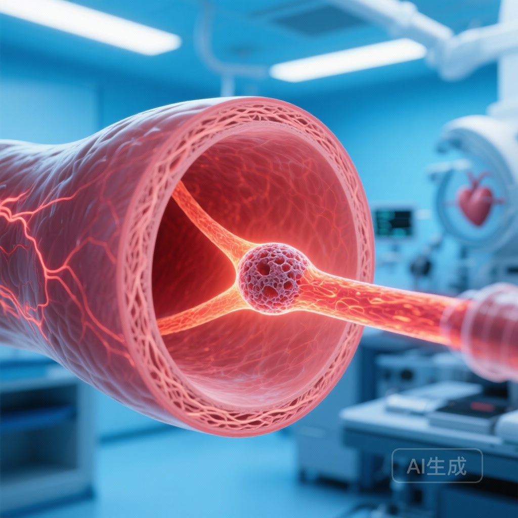
Identifying the “vulnerable plaque”—a lesion prone to rupture and subsequent thrombosis—remains a holy grail in interventional cardiology. Among various plaque morphologies, the thin-cap fibroatheroma (TCFA) has been histologically and clinically validated as the primary precursor to most acute myocardial infarctions (MI). Optical Coherence Tomography (OCT) provides the highest resolution of any intravascular imaging modality, allowing for the measurement of fibrous cap thickness and the identification of lipid-rich cores. However, the manual identification of TCFA is labor-intensive, requires specialized expertise, and is plagued by inter-observer variability.
The emergence of artificial intelligence (AI) in medical imaging offers a potential solution to these challenges. By utilizing deep learning algorithms, clinicians can potentially automate the detection of high-risk plaque features. The PECTUS-AI study was designed to evaluate whether an AI-based approach to TCFA identification could not only match but potentially enhance the prognostic utility of OCT in patients with a history of myocardial infarction.
Study Design and Methodology
The PECTUS-AI study represents a secondary analysis of the prospective, observational PECTUS-obs study. The study population consisted of 438 patients who had recently experienced a myocardial infarction and were found to have non-culprit lesions. These non-culprit lesions were deemed physiologically non-significant based on fractional flow reserve (FFR) testing (FFR > 0.80). Despite their stable physiology, these lesions were imaged using OCT to assess their morphological stability.
The researchers compared two primary methods of OCT analysis:
Manual Core Laboratory Analysis (CL-TCFA)
Independent experts manually reviewed the OCT pullbacks to identify TCFAs, defined by a fibrous cap thickness of <65 μm and a lipid arc of >90 degrees.
AI-Based Analysis (AI-TCFA)
The OCT-AID algorithm, a recently validated deep learning segmentation tool, was employed to automatically detect TCFA features across the imaged segments. The AI was tasked with evaluating both the specific “target lesion” (the FFR-negative site) and the “complete pullback” (the entire length of the artery imaged during the procedure).
The primary endpoint was a composite of all-cause mortality, non-fatal MI, or unplanned revascularization at two years, excluding events related to the initial procedure or previous stents.
Key Findings: The Superiority of AI Discrimination
The study yielded several pivotal findings that underscore the clinical utility of automated plaque analysis. Out of 414 patients with evaluable data, AI-TCFA was identified in 34.5%, while manual core lab analysis identified TCFA in 30.0%.
Prognostic Value of the Target Lesion
When focusing specifically on the target non-culprit lesion, the presence of AI-detected TCFA was significantly associated with a higher risk of the primary outcome (Hazard Ratio [HR] 1.99, 95% CI 1.02-3.90, P = .04). In contrast, the association between manually identified TCFA (CL-TCFA) and outcomes did not reach statistical significance (HR 1.67, 95% CI 0.84-3.30, P = .14). This suggests that the AI algorithm may be more sensitive or consistent in identifying features that translate into clinical risk.
The Power of the Complete Pullback
Perhaps the most striking result of the study was the prognostic power of AI when applied to the complete imaged segment. Patients with an AI-TCFA detected anywhere in the complete pullback had a significantly higher risk of experiencing the primary endpoint, with a Hazard Ratio of 5.50 (95% CI 1.94-15.62, P < .001). Furthermore, the absence of AI-TCFA in the complete pullback yielded a Negative Predictive Value (NPV) of 97.6%. This implies that if the AI does not find a TCFA in the imaged vessel, the likelihood of a major adverse cardiac event in that vessel over the next two years is exceptionally low.
Expert Commentary: Clinical Implications and Mechanistic Insights
The results of PECTUS-AI suggest a paradigm shift in how we interpret intravascular imaging. The fact that AI outperformed human experts in prognosticating events highlights the limitations of human visual assessment, which may overlook subtle but critical variations in cap thickness or lipid distribution. AI algorithms provide a level of granularity and standardization that is difficult to achieve in a busy clinical environment.
From a mechanistic standpoint, the findings reinforce the “pan-vascular” nature of atherosclerosis. The increased predictive power of the complete pullback versus the target lesion suggests that the overall burden of vulnerable plaque in a vessel is a more robust indicator of risk than a single focal point. This aligns with current understandings of systemic inflammatory drivers of plaque instability.
However, clinicians must consider the limitations. While AI identification provides strong prognostic information, the study does not yet prove that treating these AI-identified lesions (e.g., with preventive stenting or intensified medical therapy) will improve outcomes. Further randomized controlled trials, such as the ongoing PREVENT trial, will be necessary to determine if AI-guided interventions can reduce event rates.
Conclusion
The PECTUS-AI study establishes AI-based OCT analysis as a powerful tool for cardiovascular risk stratification. By providing a standardized, objective, and highly predictive assessment of thin-cap fibroatheromas, AI can assist clinicians in identifying patients at high risk for future coronary events. The high negative predictive value of full-segment AI analysis is particularly valuable, potentially allowing for a “rule-out” strategy for high-risk plaque features. As these algorithms become integrated into catheterization laboratory workflows, they promise to enhance the precision of evidence-based cardiovascular care.
Funding and ClinicalTrials.gov
The PECTUS-obs study was supported by grants from the Netherlands Organization for Health Research and Development (ZonMW). The PECTUS-AI analysis was independently conducted using the OCT-AID algorithm. ClinicalTrials.gov Identifier: NCT02852876.
References
Volleberg RHJA, Luttikholt TJ, van der Waerden RGA, et al. Artificial intelligence-based identification of thin-cap fibroatheromas and clinical outcomes: the PECTUS-AI study. Eur Heart J. 2025;46(46):5032-5041. doi:10.1093/eurheartj/ehaf595.