Introduction: Why early signs matter
Many people fear the word cancer, but cancer typically arises through a long, stepwise change from normal cells to malignant ones—often over years or decades. Before invasive cancer develops, tissues can go through recognizable precancerous stages. These early changes often cause few or no symptoms, so awareness, screening, and timely treatment are essential. In this article we explain how cancer develops, describe six common precancerous conditions, list their main risk factors, and offer practical prevention, surveillance, and treatment options.
Four stages of cancer development: a tree analogy
A useful way to picture cancer development is to imagine a tree growing from a seed:
– Seed stage: precancerous change (abnormal cell growth or tissue alteration that raises cancer risk)
– Seedling stage: carcinoma in situ (cells have become cancerous but remain confined to their original layer)
– Mature tree: invasive cancer (cancer cells penetrate surrounding tissue)
– Seed dispersal: metastatic disease (cancer spreads to distant organs)
Precancerous changes and carcinoma in situ are the best opportunities for prevention or cure. Detecting and treating abnormalities at these stages can usually prevent progression to invasive cancer.
Common precancerous conditions to watch—and what to do
Below are six precancerous states that commonly lead to specific cancers if left unchecked. For each, we describe what it is, how it is detected, major risk factors, and practical steps for prevention and management.
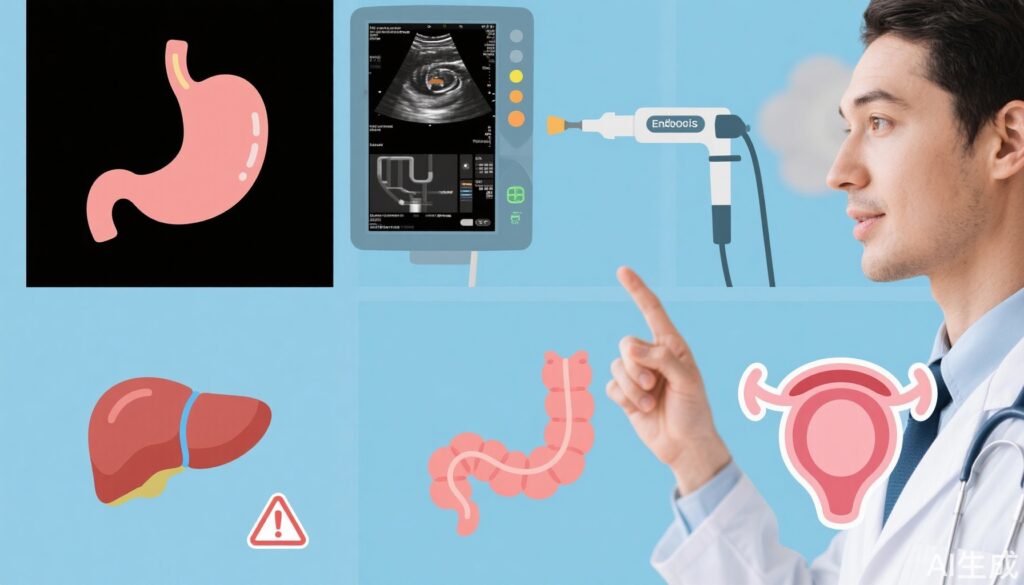
1. Chronic atrophic gastritis (stomach lining atrophy)
What it is: Chronic atrophic gastritis involves long-term thinning and loss of normal gastric glands, sometimes with intestinal metaplasia (stomach lining cells turning into intestine-like cells). This sequence can precede gastric (stomach) cancer, particularly the intestinal-type gastric adenocarcinoma.
How it’s diagnosed: Endoscopy (upper GI endoscopy) with biopsy is the diagnostic standard. Noninvasive tests such as tests for Helicobacter pylori infection and blood tests for anemia or low vitamin B12 can raise suspicion.
Main risk factors:
– Helicobacter pylori infection (classified by WHO as a carcinogen)
– High-salt and smoked or pickled foods, smoking and heavy alcohol use
– Older age and genetic predisposition
– Certain medications or bile reflux can contribute
Prevention and management:
– Eradicate H. pylori infection when present; regimens and choice of antibiotics should follow local resistance patterns and physician guidance
– Reduce salt-preserved foods, increase vegetables and fiber, stop smoking and limit alcohol
– Surveillance endoscopy and biopsy intervals are individualized depending on the severity of atrophy and presence of intestinal metaplasia; discuss with a gastroenterologist
2. Barrett’s esophagus
What it is: Barrett’s esophagus is a change of the lower esophagus lining where normal squamous cells are replaced by columnar, intestine-like cells (intestinal metaplasia). It increases the risk of esophageal adenocarcinoma.
How it’s diagnosed: Upper endoscopy with biopsy is required to confirm Barrett’s and to check for dysplasia (precancerous cellular changes).
Main risk factors:
– Long-standing gastroesophageal reflux disease (GERD)
– Obesity, especially central (abdominal) obesity
– Male sex and older age
– Smoking and a family history in some cases
Prevention and management:
– Control reflux: lifestyle measures (weight loss, avoid late meals, elevate head of bed) and proton pump inhibitors (e.g., omeprazole) to reduce acid exposure
– Endoscopic surveillance intervals depend on whether dysplasia is present; nondysplastic Barrett’s is monitored less frequently than Barrett’s with dysplasia
– Endoscopic treatments: if high-grade dysplasia or early cancer is found, endoscopic mucosal resection or radiofrequency ablation can remove or destroy abnormal tissue and sharply reduce cancer risk
– Talk with a gastroenterologist to individualize surveillance and treatment
3. Liver cirrhosis
What it is: Cirrhosis is advanced scarring of the liver with nodular regeneration and loss of normal architecture. It is the main precursor condition for primary liver cancer (hepatocellular carcinoma, HCC).
How it’s diagnosed: Diagnosis is based on imaging (ultrasound, CT, MRI), blood tests indicating liver dysfunction, and sometimes biopsy. Signs and symptoms may be absent in early compensated cirrhosis.
Main risk factors:
– Chronic viral hepatitis (hepatitis B and untreated hepatitis C)
– Alcohol-related liver disease
– Nonalcoholic fatty liver disease (related to obesity, diabetes, metabolic syndrome)
– Certain inherited metabolic disorders and toxins (e.g., aflatoxin exposure)
Prevention and management:
– Treat underlying causes: antiviral therapy for hepatitis B and curative direct-acting antivirals for hepatitis C dramatically reduce the risk of progression; lifestyle measures and weight loss for nonalcoholic fatty liver disease; abstain from alcohol
– Vaccinations: hepatitis A and B vaccines if not immune; seasonal influenza and pneumococcal vaccines as recommended
– HCC surveillance: people with cirrhosis usually undergo ultrasound of the liver every six months, often with blood tests such as alpha-fetoprotein (AFP); surveillance aims to detect tumors at a treatable stage
– Management often involves a multidisciplinary team; in advanced cases liver transplantation may be considered
4. Colorectal adenomatous polyps
What it is: Adenomatous polyps (adenomas) are benign growths of the colon or rectal lining. Over time some adenomas can progress to colorectal cancer through a sequence of genetic changes.
How it’s diagnosed: Colonoscopy is the gold standard—polyps are visualized and removed (polypectomy) and sent for histopathology. Stool-based tests and sigmoidoscopy are other screening tools but colonoscopy allows removal.
Main risk factors:
– Age (risk increases after age 50, with many programs starting screening earlier)
– Family history and genetic syndromes (e.g., familial adenomatous polyposis)
– Diets high in red/processed meat, low in fiber, obesity, smoking, and sedentary lifestyle
– Inflammatory bowel disease (ulcerative colitis, Crohn’s disease) also increases risk
Prevention and management:
– Colonoscopy with removal of adenomas prevents cancer. Surveillance intervals after polypectomy depend on number, size, and histology of removed polyps—your gastroenterologist will recommend timing (commonly 3–10 years depending on findings)
– Adopt a healthy diet rich in fiber, maintain a healthy weight, exercise, limit red/processed meats, and avoid tobacco
– Genetic counseling and earlier surveillance are indicated if there is a strong family history
5. Cervical intraepithelial neoplasia (CIN)
What it is: CIN refers to abnormal, precancerous changes of the cervical epithelium. It is graded as CIN1 (low-grade), CIN2 (moderate), and CIN3 (high-grade). High-grade lesions carry the greatest short-term risk of progressing to invasive cervical cancer.
How it’s diagnosed: Cervical screening with Pap smear and/or HPV testing identifies abnormalities. Colposcopy with directed biopsy confirms the diagnosis and grade.
Main risk factors:
– Persistent infection with high-risk human papillomavirus (HPV) types, especially HPV 16 and 18
– Early onset of sexual activity, multiple sexual partners, and other factors that increase HPV exposure
– Smoking and immunosuppression (e.g., HIV infection)
Prevention and management:
– HPV vaccination (e.g., quadrivalent or nonavalent vaccines such as Gardasil) before exposure substantially reduces the risk of CIN and cervical cancer
– Regular cervical screening per local guidelines; positive screening tests are followed by colposcopy and biopsy
– Treatment: ablative or excisional procedures such as LEEP (loop electrosurgical excision) or cold knife conization remove high-grade lesions. Follow-up is important because recurrence can occur.
6. Mucosal leukoplakia (oral and vulvar/vaginal white patches)
What it is: Leukoplakia describes white patches on mucous membranes (commonly oral, sometimes vulvar or vaginal) that cannot be scraped off and are not explained by another diagnosis. While many are benign, some contain dysplasia or will progress to squamous cell carcinoma.
How it’s diagnosed: Clinical inspection, removal of local irritants, and biopsy of suspicious or persistent lesions are used to determine whether dysplasia is present.
Main risk factors:
– Chronic irritation (dental fragments, poorly fitting dentures), tobacco, alcohol, betel nut chewing
– HPV and immunosuppression in some locations
Prevention and management:
– Remove or reduce risk factors (stop tobacco and betel nut chewing, correct dental issues)
– Biopsy any persistent, growing, or ulcerated lesion to rule out dysplasia or cancer
– For lesions with dysplasia, excision or close surveillance may be recommended
Putting it all together: prevention, screening, and when to see a doctor
Precancerous conditions are your body’s early warning signs. Key practical steps:
– Know your risk factors: family history, infections (H. pylori, hepatitis viruses, HPV), lifestyle (smoking, alcohol, diet, obesity). Reducing modifiable risks lowers chances of progression.
– Follow screening guidelines: routine screening (cervical cytology/HPV, colorectal screening, hepatitis monitoring in at-risk individuals, endoscopy when indicated) finds precancerous changes early.
– Treat underlying causes: antiviral therapy for hepatitis, H. pylori eradication, HPV vaccination, and cessation of alcohol and tobacco use are effective preventive measures.
– Don’t delay evaluation: persistent symptoms such as ongoing reflux, abnormal vaginal bleeding, oral patches, unexplained fatigue with liver disease history, or changes found on routine tests warrant timely specialist assessment.
– Discuss individualized surveillance: how often to repeat endoscopy, colonoscopy, or imaging depends on the specific lesion, pathology results, and overall risk profile.
Final note: early action saves lives
Most cancers develop slowly, and many of the precancerous stages described here can be monitored, treated, or reversed. Early diagnosis through appropriate screening and management of underlying causes is the most effective strategy to prevent progression. If you or a family member has risk factors or has been told you have one of these conditions, discuss tailored prevention and surveillance plans with your healthcare provider. That early attention can literally stop cancer in its tracks.
References and further reading: consult national screening guidelines and specialty society recommendations (gastroenterology, hepatology, gynecology, ENT/dentistry) for region-specific screening intervals and treatment protocols. Always consult a qualified clinician before starting or stopping any medical treatment.