Patient Information
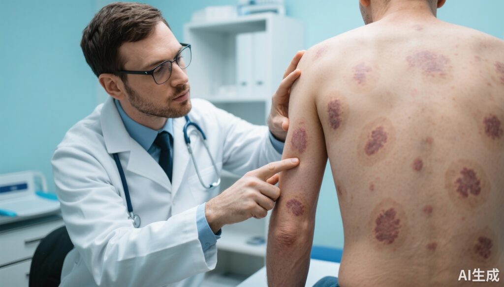
A 35-year-old female with no prior medical history presented to our dermatology clinic with multiple non-healing skin lesions involving her upper limbs and back. The lesions had progressively evolved over the past three months. She reported associated mild itching and occasional serous discharge but denied systemic symptoms such as fever, weight loss, or night sweats. There was no history of immunosuppressive conditions or exposure to individuals with active tuberculosis. The patient was immunocompetent and had no significant travel or occupational exposure relevant to her dermatologic complaints.
Diagnosis
On examination, multiple indurated plaques and nodules with erythematous edges and central ulceration were noted predominantly on her forearms and upper back. The clinical appearance suggested a chronic granulomatous infection. Skin biopsy revealed caseating granulomas with epithelioid histiocytes and Langhans giant cells. Ziehl–Neelsen staining demonstrated acid-fast bacilli within the lesions, confirming mycobacterial infection. Culture on Lowenstein-Jensen medium was positive for Mycobacterium tuberculosis complex. A chest X-ray and sputum evaluation were unremarkable, ruling out pulmonary involvement. These findings led to a diagnosis of multifocal cutaneous tuberculosis.
Differential Diagnosis
Differential diagnoses considered included:
– Cutaneous leishmaniasis: ruled out by absence of Leishmania organisms in skin smears and biopsies.
– Sporotrichosis: fungal cultures were negative; no history of trauma with plant material.
– Nontuberculous mycobacterial infections: cultures and sensitivity testing confirmed M. tuberculosis.
– Sarcoidosis: absence of systemic involvement and detection of acid-fast bacilli negated this.
– Chronic bacterial or fungal abscesses: histologic and microbiological analyses excluded other pathogens.
Treatment and Management
Standard anti-tubercular therapy (ATT) was initiated, consisting of isoniazid, rifampicin, pyrazinamide, and ethambutol during the intensive phase for two months, followed by isoniazid and rifampicin for an additional four months. The treatment regimen adhered to national tuberculosis control guidelines. The patient was monitored for adverse effects, and liver function tests were regularly assessed. Supportive wound care included gentle cleaning and topical antibiotic ointments to prevent secondary infections. The patient was counseled regarding therapy adherence and signs of toxicity.
Outcome and Prognosis
The patient demonstrated significant clinical improvement by the end of the intensive phase, with reduction in lesion size and resolution of ulcerations. At the six-month follow-up, the skin lesions had healed with minimal scarring, and no new lesions were observed. The patient remained asymptomatic with no recurrence noted at one-year post-treatment follow-up.
Discussion
Cutaneous tuberculosis is an uncommon extrapulmonary manifestation of tuberculosis, accounting for less than 2% of all TB cases. Multifocal involvement in an immunocompetent host is exceedingly rare and poses diagnostic challenges as the clinical presentation can mimic various infectious and inflammatory dermatoses. Early biopsy and microbiological confirmation are crucial for diagnosis.
This case underscores the importance of considering cutaneous TB in the differential diagnosis of chronic skin lesions, especially in endemic regions or populations at risk. The absence of systemic symptoms and pulmonary involvement should not exclude tuberculosis. Comprehensive diagnostic evaluation combining histopathology, acid-fast staining, and culture is mandatory.
Guidelines recommend standard ATT for cutaneous TB with excellent prognosis if therapy is initiated promptly. Multidisciplinary management and close follow-up are essential for treatment adherence and monitoring complications. Awareness among clinicians can help reduce misdiagnosis and delays in treatment, potentially preventing morbidity associated with delayed therapy.
References
1. Barbagallo J, Tager P, Ingleton R, Hirsch RJ, Weinberg JM. Cutaneous tuberculosis: diagnosis and treatment. Am J Clin Dermatol. 2002;3(5):319-28.
2. Frankel A, Penrose C, Emer J. Cutaneous tuberculosis: a practical case report and review for the dermatologist. J Clin Aesthet Dermatol. 2009;2(10):19-27.
3. World Health Organization. Treatment of Tuberculosis Guidelines. 4th edition. Geneva: WHO; 2010.
4. Kim YJ, Lee JS, Kim MN. Multifocal cutaneous tuberculosis in immunocompetent patient. Ann Dermatol. 2013;25(3):366-8.


