Highlight
• In a Medicare claims analysis of 19,692 BSD patients, 15% had no CT recorded within one year before the procedure (Romashko et al., JAMA Otolaryngol Head Neck Surg. 2025).
• Thirty-one point eight percent of otolaryngologists met the study’s definition of an outlier (≥10% of their BSDs without a recent CT); 6.1% of physicians accounted for >50% of missed CTs.
• Findings suggest generally good adherence to standard practice, but a meaningful minority of clinicians perform BSD without contemporary cross-sectional imaging, raising clinical, safety, and quality-monitoring concerns.
Background: Clinical context and why preoperative CT matters
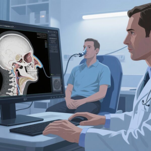
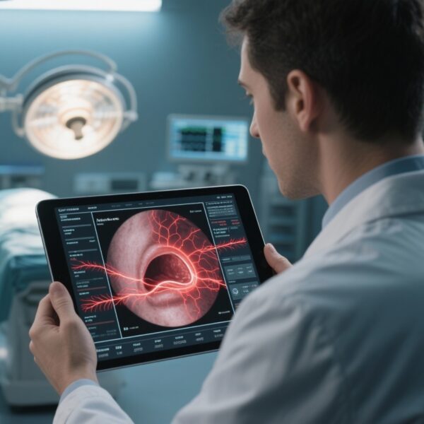
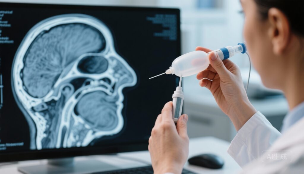
Balloon sinus dilation (BSD) has become an established option for selected patients with chronic rhinosinusitis (CRS) and recurrent acute rhinosinusitis. The procedure mechanically dilates sinus ostia using balloon catheters and is frequently performed in office settings as well as in the operating room. Preoperative cross-sectional imaging — typically noncontrast computed tomography (CT) of the paranasal sinuses — plays a central role in patient selection, procedural planning, and risk mitigation. CT provides detailed anatomic information (e.g., extent of disease, anatomic variants such as dehiscent lamina papyracea, Haller cells, or agger nasi cells, and proximity of key structures) that informs whether BSD alone is appropriate versus the need for more extensive endoscopic sinus surgery.
Most specialty guidelines and clinical practice statements endorse obtaining CT imaging when considering sinus surgery, both to confirm extent of disease and to guide operative strategy. Beyond surgical planning, contemporary imaging supports informed consent discussions and reduces the risk of complications related to unrecognized anatomic risk factors.
Study design and methods
Romashko and colleagues performed a cross-sectional analysis using Medicare claims data to evaluate adherence to practice guidance for preoperative CT before BSD. The cohort included otolaryngologists who each performed at least 11 BSD procedures between January 1, 2022, and December 31, 2023. The primary measure was the percentage of physicians who performed BSD without CT imaging available within 1 year of the procedure in >10% of their procedures; these physicians were defined as outliers. Secondary analyses explored physician-level characteristics associated with outlier status. Analyses were performed using IBM-SPSS and R.
Key findings
Population and scope
The analytic sample comprised 490 otolaryngologists (29 female, 460 male) who together performed BSD in 19,692 Medicare beneficiaries during the two-year window. The study examined claims for CT imaging up to 1 year before each BSD event.
Preoperative CT availability
Overall, 2,905 patients (15%) had no recorded CT of the sinuses within one year prior to BSD. Thus, 85% of BSDs had a documented recent CT.
Outlier physicians
One hundred fifty-six physicians (31.8%) met the study’s outlier threshold (i.e., >10% of their BSD cases lacked a prior CT). Of note, 30 physicians (6.1% of the cohort) accounted for more than 50% of all cases without preprocedure CT (1,880 patients, 9.5% of total BSDs). Another 42 physicians (8.6%) accounted for 47.5% of all missed CTs — indicating a skewed distribution in which a small subset of physicians contributed a disproportionately large share of procedures performed without recent imaging.
Interpretation of distribution
The findings suggest that while the majority of clinicians comply with expectations for preoperative imaging, nonadherence is concentrated among a minority of practitioners. The skew implies that system-level or practice-specific factors — rather than random omission — likely contribute to the absence of preprocedure CTs in many cases.
Clinical and safety implications
Patient selection and operative safety
Conducting BSD without contemporary CT limits the treating clinician’s ability to identify anatomy or disease features that might contraindicate a purely balloon-based approach or necessitate adjunctive measures. Anatomic variants (e.g., orbital dehiscence, anterior ethmoidal artery positions, or extensive disease beyond the ostium) can significantly alter risk profiles and change procedural plans.
Medico-legal and documentation concerns
From a medicolegal standpoint, lacking contemporaneous imaging may complicate defense of clinical decision-making if adverse events occur. Quality metrics and payers increasingly scrutinize adherence to standard-of-care practices; documentation of preoperative imaging is a concrete element that can be audited.
Radiation exposure and imaging alternatives
One argument sometimes offered for omitting CT is to minimize radiation exposure. However, modern low-dose and cone-beam CT protocols specifically for paranasal sinuses offer markedly reduced doses compared with routine head CT while providing diagnostic-quality detail. The tradeoff between a small additional radiation dose and the potential harms of inadequate preoperative assessment should be considered in shared decision-making.
Potential explanations for missing CTs
Data-source limitations
Claims-based analyses capture billed imaging within the payer dataset. CTs performed outside Medicare coverage (e.g., paid out-of-pocket, performed earlier than the 1-year window, or performed for non-billed reasons) may not be detected. Cross-institution imaging performed at unaffiliated centers might not appear in Medicare claims, leading to undercounting.
Practice patterns and office-based workflows
Office-based BSD programs that use in-office cone-beam CT systems may bill imaging differently or integrate imaging into facility fees, complicating capture in claims data. Conversely, some clinicians may rely on clinical history or endoscopic findings when deciding to perform BSD, particularly in patients with recurrent symptoms and known prior imaging.
Reimbursement and access barriers
Reimbursement constraints, insurance prior authorization hurdles, and limited access to timely imaging in certain geographic or resource-limited settings may prompt clinicians to proceed without a recent CT. These structural factors would be targets for policy-level solutions.
Strengths and limitations of the study
Strengths
The study leverages a large, national claims dataset covering a relevant and contemporary time interval, capturing real-world practice across many physicians. The sample size (nearly 20,000 BSD procedures) lends precision to estimates of CT utilization.
Limitations
Claims data cannot confirm whether anatomic imaging existed but was not billed to Medicare or was performed outside the 1-year window. The study lacks granular clinical detail (severity of disease, patient preferences, operative findings) that could justify deviations from guideline-based imaging. Additionally, the cohort was limited to Medicare beneficiaries, which limits generalizability to younger populations and those with other insurance arrangements.
Expert commentary and implications for practice
Clinical practice implications
The data support an overall pattern of appropriate imaging use before BSD but identify a nontrivial fraction of procedures done without recent CT. For clinicians, the takeaways are practical: obtain or confirm recent cross-sectional imaging before BSD whenever possible; document imaging review in the medical record and in informed consent discussions; and consider low-dose or cone-beam CT options to reduce radiation exposure while maintaining diagnostic quality.
Systems and policy responses
Health systems and payers should examine whether workflows, reimbursement policies, or access issues unintentionally incentivize proceeding without imaging. Quality monitoring programs could track preoperative CT rates for BSD as a measurable adherence metric. Electronic health record (EHR) alerts or order-entry checks that prompt clinicians to confirm imaging availability may reduce unintentional omissions.
Research implications
Further study is needed to link the presence or absence of preoperative CT to patient-centered outcomes (e.g., complication rates, revision surgery, symptom outcomes) and to explore why the small subset of clinicians account for most missed imaging. Qualitative work with clinicians identified as outliers would be useful to characterize barriers and beliefs driving practice.
Conclusion
Romashko et al.’s claims-based analysis provides valuable, large-scale evidence that most otolaryngologists document CT imaging before performing BSD, consistent with accepted standards. Nevertheless, the concentration of procedures done without recent imaging among a minority of physicians highlights an actionable quality gap. Addressing this gap will require combined efforts in clinician education, system-level safeguards (EHR, scheduling, and imaging access), and appropriate quality measurement to ensure safe, guideline-concordant care for patients undergoing BSD.
Funding and clinicaltrials.gov
The primary study authors reported in the JAMA Otolaryngology publication did not indicate a clinicaltrials.gov registration for this cross-sectional claims analysis. Funding sources and disclosures are reported in the original article (Romashko AA et al., 2025).
References
1. Romashko AA, Farrell NF, Kallogjeri D, Sáenz MP, Smith KA, Piccirillo JF. Preoperative Computed Tomography Utilization in Patients Undergoing Balloon Sinus Dilation. JAMA Otolaryngol Head Neck Surg. 2025 Nov 13:e254030. doi:10.1001/jamaoto.2025.4030. PMID: 41231495; PMCID: PMC12616526.
2. Rosenfeld RM, Piccirillo JF, Chandrasekhar SS, et al. Clinical practice guideline (update): Adult Sinusitis. Otolaryngol Head Neck Surg. 2015;152(2 Suppl):S1–S39. doi:10.1177/0194599815572097.
Suggested practical checklist for clinicians before BSD
• Confirm a diagnostic-quality CT of the paranasal sinuses obtained within 12 months (or sooner if symptoms changed).
• Review CT for anatomic variants or disease extent that would alter the plan.
• Document imaging review and rationale for BSD versus alternative/endoscopic approaches.
• Consider low-dose or cone-beam CT when appropriate to minimize radiation burden.
These steps can improve patient safety, support informed consent, and align practice with consensus expectations for preoperative assessment.