Patient Information
A 12-year-old female patient presented to Peking University Shenzhen Hospital in January 2020 with a chief complaint of delayed eruption of her right mandibular first molar. She reported that this dental issue had persisted for six years. The patient’s familial medical history was complex, with an undiagnosed and complicated treatment history involving the left mandible; however, no direct association with primary failure of eruption (PFE) was documented.
Facial examination revealed no notable abnormalities. Intraoral examination demonstrated partial eruption of the crown of the right mandibular first molar, with only the occlusal surface visible. The contralateral left mandibular first molar exhibited normal crown development and normal eruption. The remaining dentition displayed no eruption disturbances. Occlusal assessment showed a Class I molar and canine occlusion on the left side, while the right molars exhibited an open bite with no occlusal contact.
Dental cast analysis found mild crowding in the upper dental arch and a well-aligned lower arch. The anterior teeth presented a slight deep overbite and overjet; the curve of Spee in the lower arch measured approximately 2 mm.
Diagnosis
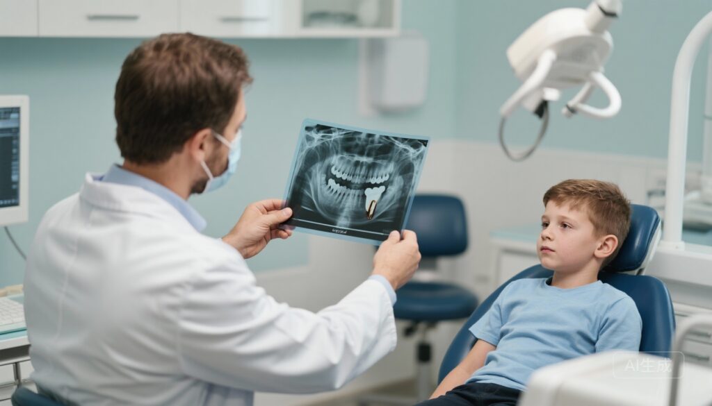
Panoramic radiography revealed the right mandibular first molar at an abnormally low vertical position, with root apices located within the mandibular canal. No significant differences in crown or root morphology were noted between the right and left first molars. The periodontal ligament space around the affected tooth was markedly narrow and varied in thickness. The lamina dura was poorly defined and discontinuous surrounding the affected molar compared with the normal left side.
A localized pit-like bone defect was observed surrounding the crown, significantly reducing mandibular bone height on the affected side. The right mandibular second premolar appeared healthy, but the right mandibular second molar was horizontally impacted, and the third molar germ showed normal development. Cephalometric analysis confirmed a Class I skeletal malocclusion.
Based on clinical, familial, and radiographic findings, the diagnosis of primary failure of eruption (PFE) affecting the right mandibular first molar was established. PFE was confirmed by the lack of response to orthodontic forces applied over two months, distinguishing it from mechanical failure of eruption.
Differential Diagnosis
The primary differential diagnoses considered and ruled out included:
– Mechanical Failure of Eruption: Ruled out due to absence of mechanical obstruction and absence of response to orthodontic force.
– Ankylosis: Excluded as radiographically the affected tooth showed a periodontal ligament space, though narrow, and no evidence of bony fusion was noted.
– Localized developmental anomalies such as odontoma or cystic lesions impeding eruption: No radiographic evidence supported these.
– Systemic causes of delayed eruption or generalized eruption failure: No systemic symptoms or involvement of other teeth were present.
Treatment and Management
The individualized treatment plan was developed considering the extent of eruption failure and patient-specific factors. The plan involved:
1. Extraction of bilateral maxillary second premolars and extraction of the affected right mandibular first molar to eliminate the non-responsive tooth.
2. Orthodontic treatment using a metal self-ligating Damon Q bracket system for alignment and retraction of the upper anterior teeth.
3. Uprighting and mesial movement of the right mandibular second molar to replace the extracted first molar, thereby aiming to establish a bilateral Class II molar occlusion.
4. Monitoring the eruption of the right mandibular third molar with potential local interventions if it could substitute for the second molar.
After two months of initial orthodontic force application, no movement of the affected molar was observed, confirming the diagnosis of PFE. Sequential arch wires were used starting with round 0.014” NiTi wires progressing to rectangular 0.019”x0.025” NiTi wires for alignment.
Rectangular 0.018”x0.025” stainless steel wires were subsequently used for leveling and space closure, combined with power chains for upper anterior teeth retraction. The right mandibular second molar was gradually uprighted using NiTi rectangular arch wires.
The total treatment duration was 34 months. After orthodontic therapy, clear dental retainers were provided for nocturnal use to ensure long-term stability.
Outcome and Prognosis
Post-treatment evaluations at 1 year demonstrated successful uprighting and mesial repositioning of the right mandibular second molar with restoration of mandibular alveolar bone integrity. The previously observed mandibular bone defect was resolved.
The patient exhibited stable occlusion with satisfactory anterior alignment and a favorable facial profile. Functionally, masticatory ability on the right side was restored. Radiographic imaging showed healthy development of the right mandibular third molar.
Routine semi-annual clinical follow-ups were scheduled to monitor long-term outcomes and to promptly address any complications.
Discussion
Primary failure of eruption (PFE) is a rare hereditary dental disorder affecting approximately 0.06% of the global population. It typically manifests as a failure of eruption in posterior permanent teeth leading to occlusal discrepancies such as open bites. PFE is characterized by a disruption of the eruption mechanism rather than obstruction by mechanical factors or ankylosis.
The pathophysiology involves abnormalities within the periodontal ligament, leading to an inability of the tooth to erupt despite an unobstructed eruption path. Affected teeth often fail to respond to orthodontic forces and may develop secondary ankylosis.
Genetic studies suggest a link between PFE and mutations in the PTH1R gene, which plays a regulatory role in bone remodeling and eruption pathways. The mutation disrupts the balance between osteogenesis and osteoclastogenesis without affecting root development, explaining the normal root morphology with failed eruption observed clinically.
Clinical diagnosis requires careful exclusion of other causes of eruption failure. Genetic testing for PTH1R mutations may aid early diagnosis but was declined in this case by the patient.
Treatment strategies vary depending on the extent of the disease. Direct restoration is generally recommended for adults, but in pediatric cases like this one, extraction of the affected tooth combined with orthodontic repositioning of adjacent teeth is preferred to restore occlusion and function.
This case highlights the importance of individualized orthodontic planning, multidisciplinary management, and long-term follow-up in managing PFE. Further research is warranted to elucidate molecular mechanisms and to optimize treatment protocols.
References
1. Thunell DH, Kornblum ND, Boaz IC, et al. Primary failure of eruption: a review and case report. J Dent Child. 2010;77(2):108-113.
2. Frazier-Bowers SA, Guo DC, Cavender A, et al. Primary failure of eruption—histopathological and genetic findings. Am J Orthod Dentofacial Orthop. 2010;137(2):198-206.
3. Sundell AL, Kurol J, Gröndahl K. Influence of initial eruption failure on subsequent tooth eruption. Eur J Orthod. 2012;34(4):419-423.
4. Guo M, Zhang W. Treatment of a mandibular molar with primary failure of eruption: A case report. Exp Ther Med. 2025;31(1):9.
5. Angle EH. Classification of Malocclusion. Dental Cosmos. 1899;41:248-264.
6. Frazier-Bowers SA. Primary Failure of Eruption: Etiology and Clinical Treatment. Semin Orthod. 2012;18(2):120-132.
7. Lee H, Kim JW, Lee KE, et al. Mutations in the PTH1R gene cause primary failure of tooth eruption. Am J Hum Genet. 2011;88(3):397-404.
8. Hendricks SJ, et al. PTH1R mutations causing primary failure of eruption: A clinical and genetic investigation. Oral Dis. 2019;25(2):570-577.
9. Smith CEL, et al. Impact of PTH1R mutations on eruption pathways in human dentition: An update. J Dent Res. 2020;99(6):694-701.


