Highlights
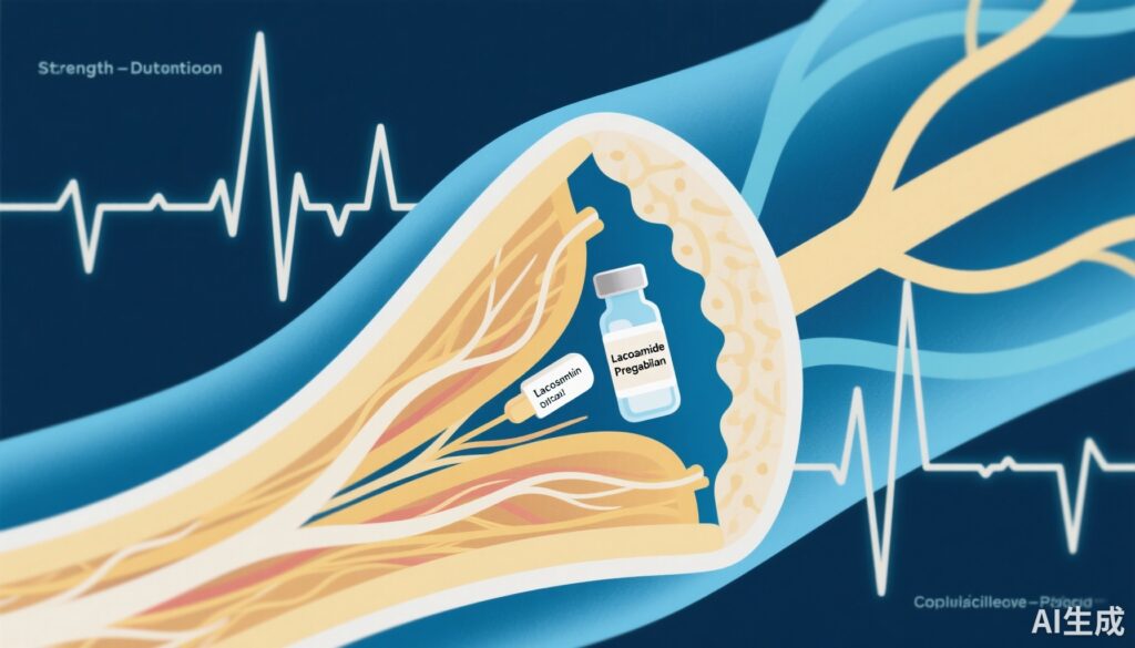
– In a randomized, double‑blind, placebo‑controlled crossover trial (n=43 healthy adults), lacosamide 200 mg acutely reduced strength–duration time constant (SDTC) in large sensory and motor fibers versus placebo at 60 minutes postdose.
– Lacosamide altered additional large‑fiber excitability parameters (subexcitability, S2 accommodation TEd measures), whereas pregabalin 150 mg and tapentadol 100 mg did not change SDTC at the same timepoint.
– The findings support nerve excitability testing as a translational pharmacodynamic biomarker for agents that modulate axonal sodium conductances, but clinical relevance for analgesia and applicability to patient populations require further study.
Background and clinical context
Chronic pain is a major cause of disability worldwide and remains difficult to treat effectively because of heterogeneous mechanisms and limited, often poorly tolerated therapeutic options. The development of analgesic drugs would benefit from validated, mechanism‑specific pharmacodynamic biomarkers that can be used early in clinical development to demonstrate target engagement and guide dose selection.
Nerve excitability testing, using threshold‑tracking techniques, interrogates axonal membrane properties noninvasively and can quantify metrics such as strength–duration time constant (SDTC), recovery cycle, and threshold electrotonus. SDTC is influenced by persistent sodium current and nodal membrane properties and therefore can serve as a readout of sodium channel modulation in peripheral axons. The IMI-PainCare-BioPain consortium aimed to standardize and validate such biomarkers; the present IMI2‑PainCare‑BioPain‑RCT1 trial tested whether lacosamide, pregabalin, and tapentadol produce measurable changes in peripheral nerve excitability in healthy volunteers.
Study design and methods
This was a randomized, double‑blind, placebo‑controlled, four‑period crossover multicenter trial in 43 healthy subjects aged 18–45 years. Each participant received single oral doses of lacosamide 200 mg, pregabalin 150 mg, tapentadol 100 mg, and placebo across four treatment periods with washout intervals. High‑frequency stimulation (HFS) was applied to induce a localized hyperalgesia model; however, the primary pharmacodynamic readouts were peripheral nerve excitability measures obtained at baseline and at prespecified postdose timepoints, with the first postdose timepoint at 60 minutes.
Two co‑primary endpoints compared lacosamide versus placebo: change in SDTC in large sensory fibers and change in SDTC in motor fibers at the first postdose assessment (60 minutes) versus baseline. Secondary predefined endpoints included components of the recovery cycle (e.g., refractoriness, relative refractory period, subexcitability), threshold electrotonus (TEd) measures, and accommodation metrics such as S2 accommodation and TEd(peak) indices. Parallel comparisons were performed for pregabalin and tapentadol versus placebo.
Key results
Primary endpoints
Lacosamide produced a statistically significant reduction in SDTC in large sensory fibers at 60 minutes postdose compared with placebo (mean reduction 0.04; 95% CI, 0.01 to 0.08; P = 0.012). A similar, statistically significant reduction was observed in motor fibers (mean reduction 0.04; 95% CI, 0.00 to 0.07; P = 0.039). These effect sizes are small in absolute units (SDTC expressed in milliseconds), but consistent across sensory and motor large myelinated fibers.
Pregabalin (150 mg) and tapentadol (100 mg) did not produce statistically significant changes in SDTC at the first postdose assessment relative to placebo.
Secondary and exploratory excitability endpoints
Among predefined secondary endpoints, lacosamide altered several large‑fiber excitability metrics: statistically significant changes were reported in subexcitability and in threshold electrotonus measures related to S2 accommodation (TEd(peak) and TEd40(Accom)). By contrast, no statistically significant differences were observed for refractoriness, relative refractory period, or accommodation half‑time at the first postdose timepoint when compared with placebo.
There was no detectable effect of lacosamide on excitability measures of small sensory fibers at the first postdose timepoint. Neither pregabalin nor tapentadol produced statistically significant changes across the majority of other predefined endpoints.
Safety and tolerability
The published report focused on pharmacodynamic endpoints in healthy volunteers; adverse event summaries are reported in the original article. Single‑dose lacosamide, pregabalin, and tapentadol in these doses are generally well characterized in healthy volunteer studies, but the present analysis emphasizes biomarker effects rather than a detailed safety evaluation in patient populations.
Interpretation and mechanistic implications
Strength–duration time constant (SDTC) is sensitive to persistent sodium conductances at the node of Ranvier and to membrane resistance. A reduction in SDTC after lacosamide is consistent with drug‑induced reduction of persistent sodium current or increased nodal membrane stabilization, reflecting lacosamide’s known mechanism as a modulator that enhances slow inactivation of voltage‑gated sodium channels. The concomitant changes in subexcitability and S2 accommodation metrics further support an effect on axonal membrane dynamics in large myelinated fibers.
Pregabalin binds to the α2δ subunit of voltage‑gated calcium channels and modulates synaptic neurotransmitter release; its primary pharmacodynamic effects are central and presynaptic rather than on peripheral axonal sodium conductance. Tapentadol combines μ‑opioid receptor agonism with noradrenaline reuptake inhibition, mechanisms that predominantly act within the central nervous system and descending pain modulatory pathways. The absence of measurable peripheral nerve excitability changes with pregabalin and tapentadol in this single‑dose healthy volunteer setting is therefore biologically plausible.
Clinical and translational relevance
These findings demonstrate that nerve excitability testing can detect pharmacodynamic modulation of peripheral axonal properties by a sodium channel‑targeting drug in humans. For early phase drug development, such objective, quantitative biomarkers can aid dose selection and proof‑of‑mechanism assessment for compounds targeting axonal excitability.
However, clinical translation from biomarker change to analgesic efficacy is not automatic. The present study used healthy volunteers and single doses; chronic dosing and disease states (e.g., diabetic neuropathy, postherpetic neuralgia) may show different patterns of axonal excitability, and the relationship between peripheral excitability changes and pain relief requires empirical validation in patient populations.
Strengths
Key strengths include randomized double‑blind crossover design, within‑subject comparisons that improve sensitivity to detect drug effects, multicenter conduct that improves generalizability, and use of standardized threshold‑tracking excitability techniques standardized by the IMI‑BioPain consortium. The trial directly compared three mechanistically distinct analgesic agents, allowing interpretation relative to their known molecular actions.
Limitations
– Healthy volunteers: The absence of neuropathic pathology limits inference about effects in patients with altered peripheral nerve function.
– Single‑dose assessment: Chronic effects, accumulation, and adaptive changes are not addressed.
– Small absolute effect sizes: While statistically significant, the clinical relevance and the relationship to analgesia remain to be determined.
– Temporal scope: The primary observation at 60 minutes postdose captures acute pharmacodynamics but may miss later or transient effects for other agents.
– Small fiber measures: Lack of lacosamide effect on small sensory fiber excitability at the early timepoint leaves open whether different techniques or later sampling would detect changes relevant to nociception.
Recommendations and future directions
To translate these biomarker findings into clinical utility, next steps should include: (1) pharmacokinetic–pharmacodynamic modeling to link plasma concentrations to excitability changes, (2) repeated‑dose studies to assess longer‑term modulation and dose–response relationships, (3) trials in neuropathic pain populations to test whether peripheral excitability changes correlate with symptom improvement, and (4) concurrent central‑and‑peripheral biomarker panels to capture multisite mechanisms of analgesic agents.
Moreover, integrating nerve excitability testing with microneurography, quantitative sensory testing, and patient‑reported outcomes could clarify which excitability changes are mechanistically and clinically meaningful for pain relief.
Conclusion
The IMI2‑PainCare‑BioPain‑RCT1 trial provides robust evidence that lacosamide acutely reduces peripheral nerve excitability in large myelinated sensory and motor fibers in healthy adults, as measured by SDTC and related excitability parameters. The absence of comparable peripheral excitability effects with pregabalin and tapentadol underscores the modality specificity of these measures. Nerve excitability testing emerges as a promising translational pharmacodynamic biomarker for agents that modulate axonal sodium conductances, but further work is required to establish links to clinical analgesia and to evaluate performance in patient populations and chronic dosing regimens.
Funding and trial registration
The study was performed within the IMI‑PainCare‑BioPain consortium (IMI2). Detailed funding sources, institutional affiliations, and trial registration identifiers are provided in the original publication: Nochi Z et al., Anesthesiology. 2025;143:1279–1295.
References
1. Nochi Z, Pia H, Bloms‑Funke P, et al. Effects of Lacosamide, Pregabalin, and Tapentadol on Peripheral Nerve Excitability: A Randomized, Double‑blind, Placebo‑controlled, Crossover, Multicenter Trial in Healthy Subjects. Anesthesiology. 2025 Nov 1;143(5):1279‑1295. doi: 10.1097/ALN.0000000000005694. PMID: 40758952; PMCID: PMC12513044.
2. Bostock H, Cikurel K, Burke D. Threshold tracking techniques in the study of human peripheral nerve. Muscle Nerve. 1998 Oct;21(10):1379‑1393. doi:10.1002/(SICI)1097-4598(199810)21:10<1379::AID-MUS6>3.0.CO;2-B. PMID: 9793931.
Author note
This article is a concise clinical and translational interpretation of the trial by Nochi et al. (Anesthesiology, 2025) intended for clinicians, clinical researchers, and health policy audiences interested in pain biomarker development and neurophysiological pharmacodynamics.