Highlight
– In a population-based cohort of 178,280 women from UK Biobank, higher baseline endogenous testosterone was associated with lower prevalence and delayed onset of knee osteoarthritis (KOA).
– Each 1 standard deviation (SD) increase in testosterone was associated with a 9% lower odds of prevalent KOA (OR 0.91; 95% CI 0.87–0.96) and a 4% longer time to incident KOA (time ratio 1.04; 95% CI 1.01–1.06) over a median 13.6-year follow-up.
– No robust associations were observed for sex hormone–binding globulin (SHBG) or estradiol after adjustment and multiple-comparison correction.
Background: Why hormones and knee osteoarthritis?
Knee osteoarthritis (KOA) is a leading cause of pain, disability, and reduced quality of life worldwide. Global burden estimates place hip and knee osteoarthritis among the most rapidly growing musculoskeletal conditions responsible for years lived with disability, especially in older adults. Women experience a higher age-adjusted prevalence of KOA than men, and risk rises sharply around the menopausal transition, suggesting a potential role for sex hormones in disease biology.
Endogenous sex steroids—principally estrogens and androgens—have biologically plausible links to joint tissues. Experimental and observational data suggest sex steroids can influence cartilage homeostasis, subchondral bone remodeling, synovial inflammation, and muscle mass and strength, all of which affect knee joint biomechanics and osteoarthritis risk. However, human epidemiologic data on circulating sex hormones and clinically meaningful KOA endpoints have been inconsistent, limited by sample size, timing of hormone measurement, and heterogeneity in outcome definition.
Study design and methods
The present analysis (Wang et al., 2025) used data from the UK Biobank, a large prospective population cohort. The investigators included 178,280 women (mean age 56.0 years) with baseline measurements of one or more of: sex hormone–binding globulin (SHBG), total testosterone, or estradiol. Outcomes were prevalent KOA at baseline and incident KOA during follow-up, with a median follow-up of 13.6 years for longitudinal analyses.
Analytic methods comprised logistic regression to estimate cross-sectional associations with prevalent KOA, and accelerated failure time (AFT) models to analyze time to incident KOA among women without baseline disease. Hormone exposures were modeled per 1 standard deviation (SD) increase and by quartiles; the authors assessed linear and nonlinear relations and adjusted for multiple comparisons. The report provides point estimates, 95% confidence intervals (CIs), and evaluates robustness across model specifications.
Key findings
Main quantitative results (reported):
– Prevalence: Higher circulating testosterone was associated with lower odds of prevalent KOA. The adjusted odds ratio was 0.91 per 1 SD increase in testosterone (95% CI 0.87–0.96), indicating a modest but statistically significant inverse association.
– Incidence: Among women free of KOA at baseline, higher baseline testosterone was associated with a longer time to incident KOA. The AFT time ratio was 1.04 per 1 SD increase in testosterone (95% CI 1.01–1.06), i.e., each SD increment in testosterone was associated with a 4% increase in event-free time over follow-up (median follow-up 13.6 years).
– Estradiol and SHBG: No consistent or statistically robust associations between baseline estradiol or SHBG and prevalent or incident KOA were observed after adjustment and multiple-comparison correction.
– Nonlinearity: The investigators did not find evidence for nonlinear (e.g., U-shaped) associations between the measured hormones and KOA outcomes after correction for multiple testing.
Interpretation of effect sizes
The observed associations, while statistically significant for testosterone, are modest in magnitude. An OR of 0.91 per 1 SD implies a small relative difference in the cross-sectional odds of KOA for women with higher versus lower testosterone within this cohort. The 4% longer time to event per SD is likewise modest but may be meaningful at a population level when accumulated over many years. Importantly, these are observational associations and cannot by themselves establish causality.
Biological plausibility and potential mechanisms
Several mechanisms could account for an inverse relation between endogenous testosterone and KOA risk in women:
- Mediator effect via muscle mass and strength: Testosterone promotes lean mass and muscle strength, which improve knee joint support and reduce abnormal loading.
- Direct effects on joint tissues: Androgens may exert chondroprotective effects through anti-inflammatory pathways or modulation of chondrocyte metabolism, although human data are limited.
- Metabolic and adipose influences: Androgens interact with adiposity and metabolic risk factors that influence OA risk; higher testosterone in women is sometimes associated with altered fat distribution and metabolic profiles.
Conversely, SHBG modulates the bioavailability of sex steroids and may confound or mediate associations, while estradiol effects are complex and vary by life stage—postmenopausal estradiol levels are low and measurement error may limit detection of associations.
Strengths
– Very large sample size with long median follow-up (13.6 years) increases statistical power to detect modest associations.
– Use of both cross-sectional and time-to-event models provides complementary perspectives on prevalent and incident disease.
– Evaluation of both linear and nonlinear relations, and consideration of multiple hormones simultaneously, helps clarify the specificity of associations.
Limitations and cautions
– Observational design: Residual confounding (for example by physical activity, prior knee injury, occupational kneeling, comorbidities, or unmeasured socioeconomic factors) may partly explain associations.
– Single baseline hormone measurement: Sex steroid concentrations vary across time and physiologic states; a single assay may misclassify long-term exposure. This is particularly relevant for estradiol, which is often near assay limits in postmenopausal women.
– Measurement and assay issues: Differences in assay sensitivity and the ability to reliably quantify low estradiol levels in older women can limit detection of true associations.
– Outcome ascertainment: The study abstract does not detail the KOA case definition (self-report, primary care data, hospital admissions, imaging-confirmed). Misclassification of disease status could bias effect estimates toward or away from the null.
– Generalizability: UK Biobank participants are volunteers and may be healthier than the general population; findings may not generalize to populations with different age distributions, ethnic composition, or health profiles.
Clinical implications
At present, these results are hypothesis-generating rather than practice-changing. The modest inverse association between endogenous testosterone and KOA risk does not warrant routine measurement of sex hormones in clinical risk stratification, nor does it support the use of testosterone replacement to prevent or treat KOA in women. Testosterone therapy carries potential adverse effects (cardiovascular, metabolic, and androgenic) and should not be used for OA prevention outside of rigorously designed clinical trials.
However, the findings highlight biologic pathways worth further study. If testosterone’s protective signal reflects improved muscle strength or anti-inflammatory effects, established interventions that increase muscle mass and function—resistance training and targeted physiotherapy—remain first-line strategies to prevent and manage KOA and may be particularly important in women with low endogenous androgens.
Research and policy recommendations
Key next steps to build on this work include:
- Mendelian randomization analyses to test for likely causal effects of lifelong higher androgen exposure on OA risk using genetic instruments for testosterone and SHBG.
- Longitudinal studies with repeated hormone measurements to reduce exposure misclassification and to model trajectories around menopause.
- Mechanistic human and animal studies that examine androgen effects on cartilage, bone, synovium, and periarticular muscle.
- Interventional trials that focus first on nonhormonal mechanisms linked to androgen status—e.g., exercise interventions to improve muscle mass and function—before considering pharmacologic androgen interventions, given safety concerns.
Conclusion
In a very large cohort of middle-aged and older women, higher endogenous testosterone was modestly associated with lower prevalence and delayed onset of knee osteoarthritis, while no robust associations were found for SHBG or estradiol. The findings are biologically plausible and suggest androgen-related pathways, possibly via muscle mass and inflammation, may influence KOA risk. Nonetheless, causality remains unproven and clinical application (for example, androgen replacement) is premature. Future causal inference studies and mechanistic research are needed to clarify whether and how sex steroids could be targeted to prevent or modify KOA in women.
Funding and clinicaltrials.gov
Funding and trial registration details are reported in the original publication (Wang J et al., Osteoarthritis Cartilage, 2025). This analysis used UK Biobank observational data and was not an interventional trial registered on clinicaltrials.gov.
References
1. Wang J, Yin J, Zhang X, Wang J, Xing X, Tu J, Cai G. The association between endogenous sex hormones and knee osteoarthritis in women: A population-based cohort study. Osteoarthritis Cartilage. 2025 Oct;33(10):1229-1236. doi: 10.1016/j.joca.2025.02.782. PMID: 40058626.
2. Cross M, Smith E, Hoy D, et al. The global burden of hip and knee osteoarthritis: estimates from the Global Burden of Disease 2010 study. Ann Rheum Dis. 2014 Jul;73(7):1323-1330. doi:10.1136/annrheumdis-2013-204763.
3. Felson DT. Osteoarthritis of the knee. N Engl J Med. 2006 Feb 23;354(8):841-848. doi:10.1056/NEJMcp052771.
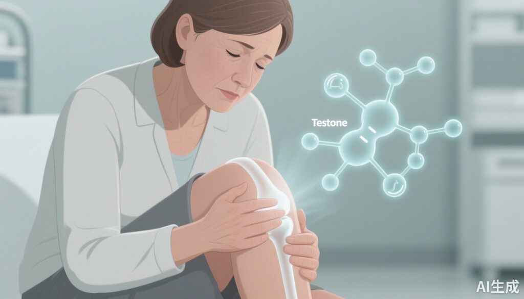
AI image prompt for article thumbnail
A realistic, clinical-style image of a middle-aged woman seated in a neutral-toned examination room, one hand gently supporting her knee in mild discomfort. Superimposed translucent medical overlays show a stylized knee joint and schematic hormone molecules (hexagonal chemical icons labeled “T” for testosterone). Lighting is soft and professional; color palette is muted blues and greys with a hint of warm skin tones; composition centers on the patient’s face and knee to convey human impact and scientific inquiry.