Highlights
- Prospective cohort study links prenatal chlorpyrifos exposure with widespread cortical thickness alterations and reduced white matter volumes in children aged 6 to 15.
- Multi-modal MRI modalities (anatomic, spectroscopic, diffusion, and perfusion imaging) reveal consistent dose-dependent brain structure and metabolism disturbances.
- Neurotoxicity is potentially mediated through inflammation and oxidative stress pathways, suggesting common mechanistic pathways with other prenatal environmental exposures.
- Policy implications emphasize the importance of minimizing prenatal pesticide exposure to protect neurodevelopment.
Background
Chlorpyrifos (CPF) is a globally prevalent chlorinated organophosphate pesticide widely used in agriculture and indoor pest control until regulatory restrictions began in the early 2000s due to mounting evidence of neurotoxicity. The prenatal period is especially vulnerable to environmental toxins because it encompasses critical stages of brain development, including neuronal proliferation, migration, differentiation, and myelination. Disruptions during this window may lead to persistent neurodevelopmental deficits.
To date, preclinical animal models and epidemiologic studies have flagged CPF as neurotoxic, but direct correlations with brain morphology in humans remained uncharacterized until the referenced prospective longitudinal study published in JAMA Neurology (Peterson et al., 2025).
Key Content
Study Design and Population
The study longitudinally followed 727 pregnant African American and Dominican women from Northern Manhattan and the South Bronx recruited from 1998 to 2006, an inner-city demographic with documented high prenatal pesticide exposure primarily through indoor residential use prior to 2001 bans. MRI data were obtained from 332 children aged 6-14.7 years between 2007 and 2015, with final analyses on 270 children with both imaging and prenatal chlorpyrifos biomarker data.
Imaging Modalities and Findings
The investigation incorporated multi-modal MR techniques:
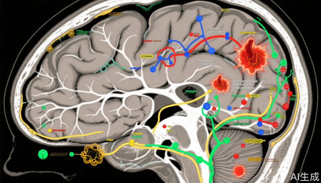
- Anatomic MRI: Demonstrated thicker cortices in frontal (superior, middle, inferior frontal gyri, anterior cingulate, orbitofrontal cortex), temporal (superior, middle, inferior temporal gyri, parahippocampus), and posterior inferior regions (cingulate cortex, cuneus, fusiform gyrus) with higher CPF exposure. Conversely, reduced cortical thickness occurred in dorsal parietal (superior parietal gyrus).
- White Matter Measures: CPF exposure correlated with decreased white matter volumes in multiple lobes and reduced diffusivity metrics in internal capsule fibers.
- Magnetic Resonance Spectroscopic Imaging (MRSI): Revealed inverse correlations between CPF levels and N-acetyl-L-aspartate (NAA), a neuronal integrity marker, particularly in deep white matter and insular cortex regions.
- Diffusion Tensor Imaging (DTI): Revealed higher fractional anisotropy and lower average diffusion coefficient in internal capsule associated with CPF exposure, indicating altered white matter microstructure.
- Arterial Spin Labeling (ASL): Showed reduced regional cerebral blood flow with increasing CPF exposure.
These imaging biomarkers collectively suggest CPF exposure disrupts not only brain structure but also neurochemical and hemodynamic aspects critical for normal development.
Neurodevelopmental and Functional Correlates
Prenatal CPF levels linked to diminished neuronal density in white matter tracts corresponded with poorer fine motor skills and motor programming in children — outcomes aligned with observed imaging abnormalities.
Mechanisms and Shared Toxicologic Pathways
Despite CPF’s distinct chemical nature, observed neuroimaging signatures paralleled those seen in prenatal exposures to air pollution, suggesting common pathophysiology principally driven by neuroinflammation and oxidative stress targeting developing neurons.
Population and Public Health Considerations
The studied population highlights disproportionate exposure risks to urban, minority communities with indoor pesticide use and proximity to agricultural pesticide drift. Elevated fetal tissue burdens reflect CPF’s placental transfer and capacity to traverse the immature fetal blood-brain barrier.
Expert Commentary
Dr. Bradley S. Peterson emphasized the robustness of the dose-response relationship between prenatal CPF exposure and various brain metrics and motor deficits, marking a landmark in human in vivo confirmation of CPF’s neurotoxicity. The convergence of structural, metabolic, diffusion, and perfusion abnormalities across brain regions implies complex and multifaceted impact on neurodevelopment.
Limitations include observational design and residual confounding; however, longitudinal design and multimodal imaging strengthen causal inference. Research gaps remain in therapeutic mitigation for exposed children—current strategies focus exclusively on primary prevention through exposure reduction.
Regulatory shifts such as the 2021 EPA tolerance revocation underscore policy responsiveness, though legal challenges reflect ongoing controversy surrounding pesticide use balancing agricultural economics and public health.
Conclusion and Future Directions
This comprehensive human study substantiates that prenatal chlorpyrifos exposure has lasting, dose-dependent detrimental effects on brain structure, neuronal integrity, and motor function detectable well into mid-childhood. These findings warrant stringent policies to restrict CPF exposure prenatally and highlight the critical need for public health education targeting at-risk populations.
Further studies should elucidate the reversibility of CPF-induced neurotoxicity, explore interventions to attenuate oxidative and inflammatory damage, and identify biomarkers predictive of neurodevelopmental outcomes. The integration of neuroimaging with functional assessments provides a translational framework to link environmental exposures to neurobiological endpoints.
References
- Peterson BS, Delavari S, Bansal R, et al. Brain Abnormalities in Children Exposed Prenatally to the Pesticide Chlorpyrifos. JAMA Neurol. 2025 Aug 18. doi:10.1001/jamaneurol.2025.2818. Epub ahead of print. PMID: 40824645.
- Rauh VA, Arunajadai S, Horton M, et al. 7-Year neurodevelopmental scores and prenatal exposure to chlorpyrifos, a common agricultural pesticide. Environ Health Perspect. 2011;119(8):1196-1201. doi:10.1289/ehp.1003160
- Eskenazi B, Marks AR, Bradman A, et al. Organophosphate pesticide exposure and neurodevelopment in young Mexican-American children. Environ Health Perspect. 2007;115(5):792-798. doi:10.1289/ehp.9828


