Patient Information
Two male siblings, aged 14 years (elder) and 12 years (younger), born to a consanguineous couple, were referred to the Department of Pedodontics and Preventive Dentistry with the chief complaint of decayed teeth. Both had been diagnosed at birth with lamellar ichthyosis (LI) and had long-standing dermatologic care consisting of topical emollients and supportive ophthalmic therapy for photophobia and ectropion-related symptoms. No other systemic abnormalities were reported.
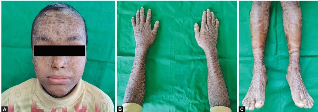
On extraoral examination both boys demonstrated the typical cutaneous phenotype of LI: thick, pigmented, adherent, armor-like scales distributed over the face, scalp, trunk, and extremities, with sparse and short scalp hair. Both had a history of presenting as collodion babies at birth and reported hypohidrosis and photosensitivity. No nail dystrophies were noted. A three-generation pedigree documented affected siblings and consanguinity in the parents.
Intraoral findings differed slightly between the siblings. The elder (14-year-old) had a full permanent dentition with erupting premolars and enamel defects localized to the occlusal surfaces of permanent maxillary and mandibular molars; dental caries was present on tooth 16. The younger (12-year-old) was in the mixed dentition stage and exhibited enamel defects involving primary and permanent molars with carious lesions on 55, 65, 26, 36 and 46. The affected teeth were sensitive to cold. There was no clinical evidence of periodontal destruction or gross alteration in salivary flow in either patient.
Diagnosis
Key clinical features supporting the diagnosis:
– Typical dermatologic phenotype of lamellar ichthyosis: collodion membrane at birth followed by generalized hyperkeratotic, plate-like scales and ectropion/photophobia. (History and examination as documented.)
– Family history consistent with autosomal recessive inheritance and parental consanguinity.
Although genetic testing results were not available in these cases, the clinical diagnosis of LI was established based on the neonatal collodion presentation, characteristic scaling, and family history. The presence of enamel hypoplasia/defects with increased caries susceptibility was diagnosed clinically by visual and tactile dental examination.
Differential Diagnosis
The following conditions were considered and excluded based on the clinical presentation and history:
– Nonbullous congenital ichthyosiform erythroderma (NBCIE): NBCIE may present with erythroderma and fine scaling rather than the large, plate-like scales typical of LI; neonatal collodion membrane is more characteristic of LI [7,8].
– Epidermolytic ichthyosis (formerly bullous ichthyosiform erythroderma): Characteristically presents with blistering and widespread erythroderma; clinical history lacked blistering episodes [3,8].
– Syndromic ichthyoses (e.g., Netherton syndrome, Sjögren–Larsson syndrome): These have distinctive systemic features (hair shaft abnormalities, neurologic impairment, ichthyosis with intellectual disability, etc.) not present in these siblings [3].
– Secondary enamel defects from systemic causes (e.g., congenital infections, nutritional deficiencies): Vitamin D levels were reported as normal in these patients and there was no history of perinatal infection or metabolic disease; the distribution of enamel defects in molars and associated caries were compatible with enamel hypoplasia related to the underlying genetic disorder described in prior reports [2–6].
Treatment and Management
Dermatologic care:
– Both patients continued routine dermatologic management with topical emollients to maintain skin hydration and to reduce scaling. Ophthalmic lubricants/eye drops were used for symptomatic relief of dryness and photophobia. Protective tinted eyewear was provided during dental procedures to mitigate photophobia and ocular discomfort.
Dental management:
– Prophylactic oral hygiene reinforcement and education, including supervised toothbrushing recommendations and use of remineralizing toothpaste, were provided to both siblings.
– Restorative care: Carious lesions were restored following standard operative protocols appropriate for patient age and tooth vitality. The elder sibling received restoration of tooth 16; the younger sibling received restorations for primary molars (55, 65) and permanent molars (26, 36, 46) as indicated. Care was taken when manipulating perioral tissues because affected skin areas may be tender or friable.
– Sensitivity management: For teeth with sensitivity, desensitizing agents and remineralization strategies were used.
– Infection control and procedural planning: Given potential dermatologic fragility and decreased barrier function of the skin, gentle handling and avoidance of excessive mechanical trauma were emphasized.
– Medication considerations: The dental team reviewed the patients’ systemic medications with their dermatologist. Although topical retinoids have limited systemic absorption, patients receiving systemic retinoids require caution due to potential hepatotoxicity; when present, hepatic considerations may influence selection and dosing of local anesthetic agents or systemic antibiotics. In these siblings, no systemic retinoid therapy was reported at presentation, but the dental team remained alert to medication interactions and hepatic risk [4,11].
– Follow-up: Both patients were scheduled for recall visits at three-month intervals for prophylaxis, reinforcement of oral hygiene, monitoring of restorations and eruption of developing teeth.
Outcome and Prognosis
Both siblings underwent successful dental prophylaxis and restorative treatment without intraoperative complications. Protective eyewear reduced photophobic discomfort during treatment sessions. Regular follow-up visits were planned at three-month intervals for preventive care and monitoring. No periodontal destruction, major salivary dysfunction, or progressive dental complications were noted during the initial follow-up period described.
Long-term prognosis for oral health in LI patients depends on early preventive care, management of enamel defects, and control of caries risk factors. There is no cure for LI; cutaneous and ophthalmic symptoms are managed symptomatically. With ongoing dermatologic and dental care, patients can maintain acceptable oral health, although lifelong vigilance is required due to the risks associated with enamel defects and increased caries susceptibility.
Discussion
Lamellar ichthyosis is a rare autosomal recessive disorder of keratinization most commonly associated with mutations in the TGM1 gene, leading to defective formation of the cornified cell envelope and impaired skin barrier function [4,5]. The reported incidence is low (<1:300,000), and LI often presents as a collodion baby at birth, later developing the characteristic plate-like scales [1,7]. Typical extracutaneous features include ectropion, hypohidrosis, photosensitivity, and nail dystrophies in some cases [1,8]. Parental consanguinity increases the likelihood of biallelic pathogenic variants and is a commonly reported history [5].
The oral manifestations of LI are less well characterized in the literature. Case reports and small series have described a spectrum of findings including enamel hypoplasia/defects, premature tooth loss, gingival hyperplasia, reduced mouth opening, tooth crowding, retained primary teeth, and increased caries prevalence [2–4,6,10]. The two siblings presented here primarily exhibited enamel defects affecting molars with associated dental caries and dentinal sensitivity—findings congruent with previously reported cases [4,6,10]. In contrast to some published reports, neither sibling demonstrated hyperplastic gingival tissue, altered salivary flow, nor periodontal destruction, and vitamin D levels were normal in these patients [2,4].
Mechanisms linking LI and dental enamel defects are not fully elucidated. Possible explanations include shared pathways of epithelial differentiation during tooth development, genetic influences on enamel matrix formation, and increased susceptibility to perinatal or postnatal environmental insults due to compromised barrier functions [4]. Enamel hypoplasia predisposes to early caries development by creating niches for plaque accumulation and weakening the enamel surface.
Dental management of patients with LI requires multidisciplinary coordination with dermatology and ophthalmology. Practical considerations include:
– Gentle handling of fragile perioral skin to avoid fissuring or trauma.
– Photophobia precautions (tinted eyewear) during dental procedures.
– Review of systemic medications, particularly systemic retinoids, due to potential hepatic toxicity and drug interactions that may affect choices of local anesthetics, antibiotics, or other systemic agents [4,11].
– Emphasis on preventive regimens: topical fluoride, casein phosphopeptide–amorphous calcium phosphate (CPP-ACP) or other remineralizing agents, dietary counseling, and frequent recall intervals to monitor restorations and reinforce oral hygiene.
These cases underscore the importance of early dental assessment for children with LI. Timely preventive and restorative care can mitigate caries progression and help preserve dentition. Clinicians should maintain a high index of suspicion for enamel defects and plan long-term individualized preventive programs.
Limitations: Genetic testing to confirm pathogenic variants (e.g., TGM1 mutation) was not reported for these patients; however, the clinical presentation and family history were strongly supportive of LI. Longer-term dental follow-up data were not available in the report.
References
1. Huang JJ, Huang MY, Huang TY. Lamellar ichthyosis with severe bilateral ectropion and self-healing collodion membrane. Biomark Genomic Med 2013;5(3):110–112.
2. Tewari N, Mathur VP, Tamchos R, et al. Oral manifestations of lamellar ichthyosis in association with rickets. BMJ Case Rep 2020;13(7):e235008.
3. Oji V. Clinical presentation and etiology of ichthyoses. Overview of the new nomenclature and classification. Hautarzt Z Dermatol Venerol Verwandte Geb 2010;61(10):891–902; quiz 903–904.
4. Nair KK, Kodhandram GS. Oral manifestations of lamellar ichthyosis: a rare case report. Indian J Paediatr Dermatol 2016;17(4):283–286.
5. Youssefian L, Vahidnezhad H, Saeidian AH, et al. Autosomal recessive congenital ichthyosis: genomic landscape and phenotypic spectrum in a cohort of 125 consanguineous families. Hum Mutat 2019;40(3):288–298.
6. Rathi NV, Rawlani SM, Hotwani KR. Oral manifestations of lamellar ichthyosis: a rare case report and review. J Pak Assoc Dermatol 2013;23(1):99–102.
7. Akiyama M, Sawamura D, Shimizu H. The clinical spectrum of nonbullous congenital ichthyosiform erythroderma and lamellar ichthyosis. Clin Exp Dermatol 2003;28(3):235–240.
8. Oji V, Traupe H. Ichthyoses: differential diagnosis and molecular genetics. Eur J Dermatol 2006;16(4):349–358.
9. Craiglow BG. Ichthyosis in the newborn. Semin Perinatol 2013;37(1):26–31.
10. Pranitha V, Thimma Reddy BV, Daneswari V, et al. Lamellar icthyosis–a case report. J Clin Diagn Res 2014;8(11):ZD01.
11. Sandberg NO. Lamellar ichthyosis. Pediatr Rev 1981;2(7):213–216.
12. Martin MD, Nusbacher C. The patient with ichthyosis. Report of a case. Oral Surg Oral Med Oral Pathol 1985;59(6):581–584.
Author note
This report is based on the published case series by D’Souza et al.: “Oral Manifestations of Lamellar Ichthyosis: A Case Report of Two Siblings” (Int J Clin Pediatr Dent. 2025 May;18(5):606-609). The present structured case report emphasizes dental implications, management considerations, and multidisciplinary coordination in patients with lamellar ichthyosis.