Highlight
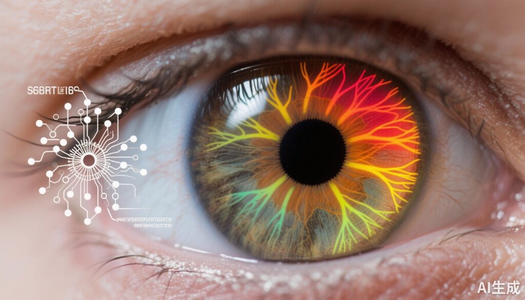
• A deep learning (machine-to-machine) model can accurately predict retinal nerve fiber layer (RNFL) thickness from optic disc photographs in ocular hypertension patients.
• Lower baseline predicted RNFL thickness significantly correlates with higher risk of developing primary open-angle glaucoma (POAG).
• Longitudinal decline in predicted RNFL thickness strongly predicts conversion to POAG, enhancing glaucoma risk monitoring.
• Incorporation of predicted RNFL thickness complements established clinical risk factors to improve risk stratification in glaucoma management.
Study Background and Disease Burden
Primary open-angle glaucoma (POAG) is a leading cause of irreversible blindness worldwide, characterized by progressive retinal ganglion cell loss and optic nerve damage. Elevated intraocular pressure (ocular hypertension) is a major modifiable risk factor for POAG, yet not all individuals with ocular hypertension develop glaucoma. Accurate identification of patients at highest risk for glaucoma conversion remains a critical challenge to prevent vision loss through timely intervention.
Retinal nerve fiber layer (RNFL) thickness, measurable by optical coherence tomography (OCT), is a sensitive biomarker for early glaucomatous damage. However, OCT devices may not be widely accessible in all clinical settings, and longitudinal OCT imaging is resource-intensive. Conversely, optic disc photography is a widely available diagnostic tool. Harnessing deep learning to predict RNFL thickness from optic disc photographs could provide a scalable, cost-effective strategy to assess glaucoma progression risk.
Study Design
This diagnostic study leveraged data from the landmark Ocular Hypertension Treatment Study (OHTS) 1 and 2 trials, enrolling 1,636 participants (3,272 eyes) with ocular hypertension but no glaucoma at baseline. The multi-center OHTS trials spanned from 1994 to 2008, providing long-term follow-up and comprehensive clinical data.
The study utilized 66,714 optic disc photographs obtained longitudinally from this cohort. A machine-to-machine (M2M) deep learning model, previously trained on OCT-derived RNFL thickness measurements, was applied to predict RNFL thickness from each photograph. Predicted baseline RNFL and longitudinal changes were analyzed alongside demographic and clinical variables.
Primary outcomes included assessing predicted RNFL thickness as an independent risk factor for conversion from ocular hypertension to POAG, using Cox proportional hazards models in univariable and multivariable frameworks.
Key Findings
Among 1,444 analyzed participants, average age at baseline was 56 years, with 57.7% female. Key discoveries include:
- Mean baseline predicted RNFL thickness was significantly thinner in eyes that eventually converted to POAG (94.1 µm) compared to those that did not (97.1 µm), indicating a 3.0 µm mean difference (95% CI, 2.2–3.8; P < .001).
- In univariable analysis, each 10-µm decrement in predicted RNFL thickness nearly doubled the hazard of glaucoma conversion (HR 1.97; 95% CI, 1.60–2.42; P < .001).
- This association remained robust after adjusting for age, intraocular pressure, central corneal thickness, visual field pattern standard deviation, mean deviation, and cup-disc ratio in multivariable models (HR 1.83; 95% CI, 1.49–2.25; P < .001).
- Longitudinally, faster loss in predicted RNFL thickness (per 1 µm/year) was a powerful predictor of conversion (HR 6.01; 95% CI, 3.33–10.64; P < .001), highlighting its utility for disease progression monitoring.
These findings indicate that deep learning-predicted RNFL thickness from standard optic disc photographs serves as a sensitive biomarker for both baseline glaucoma risk and dynamic disease progression in patients with ocular hypertension.
Expert Commentary
The study by Liu et al. represents a significant advancement in glaucoma diagnostics by integrating artificial intelligence (AI) with traditional imaging. Deep learning-based RNFL prediction facilitates risk assessment in settings lacking OCT technology, potentially democratizing early glaucoma detection globally.
Importantly, the M2M model’s reliance on optic disc photographs, a widespread and accessible modality, ensures broad applicability of this approach. The strong independent association between predicted RNFL thinning and glaucoma conversion underscores its biological plausibility given RNFL’s role in retinal ganglion cell integrity.
However, some limitations warrant attention. The original training data and validation were confined to OHTS populations primarily of certain ethnicities and clinical contexts; external validation in diverse populations and real-world clinical settings is essential. The retrospective study design and image quality variability could influence model performance.
Future prospective studies may integrate predicted RNFL thickness with other AI-based biomarkers and multimodal imaging to refine personalized glaucoma risk prediction and management strategies.
Conclusion
The application of an OCT-trained deep learning model to optic disc photographs enables accurate prediction of RNFL thickness, providing a non-invasive, accessible biomarker of glaucoma risk in ocular hypertension. Both baseline predicted RNFL thickness and longitudinal thinning rates portend increased risk of converting to primary open-angle glaucoma.
Incorporation of predicted RNFL thickness into clinical workflows could enhance early identification and monitoring of patients at highest risk, informing timely therapeutic interventions. Further research validating this approach across varied populations and integrating it into decision support systems may facilitate improved glaucoma outcomes worldwide.
References
1. Liu JC, Jammal AA, Scherer R, da Costa DR, Kass M, Gordon M, Medeiros FA. Predicting Retinal Nerve Fiber Layer Thickness From Ocular Hypertension Treatment Study Optic Disc Photographs. JAMA Ophthalmol. 2025 Aug 1;143(8):652-659. doi:10.1001/jamaophthalmol.2025.1740. PMID: 40569586; PMCID: PMC12203391.
2. Kass MA, Heuer DK, Higginbotham EJ, et al. The Ocular Hypertension Treatment Study: a randomized trial determines that topical ocular hypotensive medication delays or prevents the onset of primary open-angle glaucoma. Arch Ophthalmol. 2002;120(6):701-713.
3. Weinreb RN, Aung T, Medeiros FA. The pathophysiology and treatment of glaucoma: a review. JAMA. 2014;311(18):1901-1911.
4. De Moraes CGV, Liebmann JM, Levin LA. Glaucoma. Lancet. 2021;397(10280):2508-2521.


