Highlight
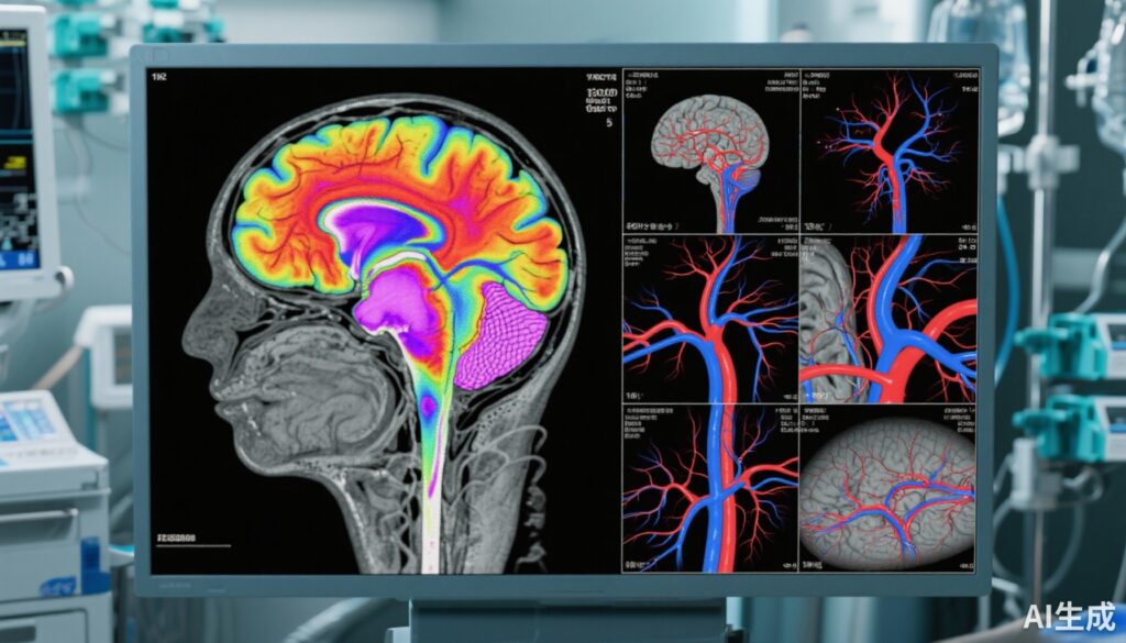
- Qualitative brainstem CT perfusion demonstrated high sensitivity (98.5%) but moderate specificity (74.4%) for diagnosing death by neurologic criteria (DNC).
- Whole-brain CT perfusion showed balanced sensitivity (93.6%) and specificity (92.3%), outperforming CT angiography in specificity.
- CT angiography showed variable sensitivity (75.5%–87.3%) and high specificity (approximately 90%), with excellent interrater reliability across imaging modalities.
- No serious adverse events were reported; minor, transient events occurred in 5% of patients.
Study Background
Confirmation of death by neurologic criteria (DNC), commonly referred to as brain death, is a critical clinical milestone to guide patient management and facilitate organ donation. Clinical examination remains the cornerstone of DNC determination; however, ancillary tests are often required when parts of the physical examination are confounded or incomplete. Current ancillary tests such as electroencephalography or radionuclide cerebral blood flow studies have limitations related to diagnostic accuracy, availability, and validation. Computed tomography (CT)-based techniques, including CT perfusion and CT angiography, offer rapid and widely accessible options for assessing cerebral blood flow and vascular patency. However, prior to this large multicenter evaluation, their diagnostic accuracy and safety profiles for DNC were inadequately defined.
The study by Chassé et al. addresses this gap by rigorously assessing the diagnostic value of CT perfusion and CT angiography as ancillary tests for brain death confirmation in a real-world critical care setting.
Study Design
This prospective, blinded diagnostic accuracy study took place from April 2017 to March 2021 across 15 adult intensive care units throughout Canada, spanning a diverse patient population. Inclusion criteria comprised critically ill adults aged 18 years or older, all with a Glasgow Coma Scale (GCS) score of 3 (indicating deep coma or unresponsiveness) and no confounding factors that could affect neurological examination or death determination.
Within two hours of a standardized clinical DNC examination, patients underwent contrast-enhanced brain CT perfusion with CT angiographic reconstruction. Two independent neuroradiologists, blinded to clinical data and each other’s assessment, interpreted imaging qualitatively and quantitatively.
The primary endpoint was the diagnostic sensitivity and specificity of brainstem CT perfusion for DNC. Secondary assessments included whole-brain CT perfusion, CT angiography performance, interrater reliability using Cohen’s kappa coefficient, and safety regarding imaging-related adverse events.
Key Findings
A total of 282 patients (mean age 57.8 years; 47% female) completed the full study protocol. Of these, 204 patients (72%) were declared dead by standardized clinical criteria.
Diagnostic Performance
- Brainstem CT Perfusion (qualitative): Showed excellent sensitivity at 98.5% (95% CI 95.8%-99.7%), meaning it correctly identified almost all patients with DNC. However, specificity was moderate at 74.4% (95% CI 63.2%-83.6%), indicating a notable false-positive rate.
- Brainstem CT Perfusion (quantitative): Did not achieve diagnostic accuracy sufficient for clinical use, suggesting qualitative interpretation remains preferred.
- Whole-brain CT Perfusion (qualitative): Demonstrated a strong balance with sensitivity of 93.6% (89.3%-96.6%) and specificity of 92.3% (84.0%-97.1%). This suggests whole-brain assessment may provide superior overall diagnostic reliability than brainstem-only evaluation.
- CT Angiography: Sensitivity ranged from 75.5% (95% CI 69.0%-81.2%) to 87.3% (81.9%-91.5%), with specificity between 89.7% and 91.0%. Although specificity was high, CT angiography was less sensitive than perfusion studies.
Interrater Reliability and Safety
The blinded neuroradiologists showed excellent agreement for all imaging modalities with Cohen κ values ranging from 0.81 to 0.84, indicating strong reproducibility.
Regarding safety, 14 patients (5%) experienced minor, self-limited adverse events such as mild contrast reactions or transient physiological changes; importantly, no serious adverse events occurred related to the imaging studies.
Expert Commentary
The findings confirm that contrast-enhanced brain CT perfusion, particularly qualitative whole-brain assessment, can reliably support the diagnosis of DNC with high sensitivity and specificity, though neither CT perfusion nor CT angiography reached the prespecified validation threshold of >98% specificity necessary to supplant clinical diagnostic criteria.
This underscores that ancillary imaging serves as a valuable but adjunctive tool rather than a stand-alone test.
Notably, the moderate specificity of brainstem CT perfusion highlights risks of false-positive diagnosis if used in isolation, potentially leading to premature or erroneous declarations of brain death. The superior specificity of whole-brain perfusion may be explained by capturing more global cerebral blood flow dynamics rather than localizing solely to brainstem perfusion.
Considering the safety profile and excellent interrater reliability, these modalities offer practical ancillary testing options in intensive care, particularly where traditional tests are less feasible.
Limitations include exclusion of confounding clinical states limiting generalizability, and the study’s single-country setting. Future research may explore combining imaging modalities or integrating advanced machine learning techniques to enhance diagnostic accuracy.
Conclusion
CT perfusion and CT angiography offer promising ancillary investigations for the assessment of death by neurologic criteria with high sensitivity and acceptable safety. However, due to suboptimal specificity and diagnostic performance below the rigorous validation threshold, these modalities should complement—rather than replace—comprehensive clinical neurological examination. This approach ensures robust, timely confirmation of brain death critical for ethical patient care and organ donation protocols.
Clinicians should interpret ancillary CT imaging cautiously within the broader clinical context, integrating results with standardized exams and other ancillary tests as necessary. Continued multidisciplinary research is needed to optimize brain death diagnostic pathways and incorporate evolving neuroimaging technologies reliably into clinical practice.
Funding and Clinical Trials Registration
This study was supported by the Canadian Institutes of Health Research and the Canadian Critical Care Trials Group. The protocol is registered with ClinicalTrials.gov (identifier: NCT03232322).
References
1. Chassé M, Shankar JJS, Fergusson DA, et al. Computed Tomography Perfusion and Angiography for Death by Neurologic Criteria. JAMA Neurol. 2025;82(9):932-940. doi:10.1001/jamaneurol.2025.2375.
2. Wijdicks EF. The Diagnosis of Brain Death. N Engl J Med. 2001;344(16):1215–1221. doi:10.1056/NEJM200104193441607.
3. Shemie SD, Doig C, Dickens B, et al. Death Determination and Organ Donation: A Model Policy for Organ Donation Organizations. Can J Anaesth. 2020;67(12):1851-1862. doi:10.1007/s12630-020-01842-7.
4. Greer DM, Varelas PN, Haque S, et al. Variability of Brain Death Policies in the United States. Neurology. 2016;86(14):1254-1259. doi:10.1212/WNL.0000000000002507.