Highlights
– Continuous intravenous sedation in early acute hypoxemic respiratory failure (AHRF) produces electroencephalographic (EEG) patterns—termed EEG Ups—not observed in natural sleep.
– EEG Ups occupied a large portion of monitored time (42% overall; >50% with some sedative–opioid regimens) and showed dose- and drug-dependent associations.
– EEG Ups were associated with deeper clinical sedation scores and, importantly, with higher ICU mortality.
Background
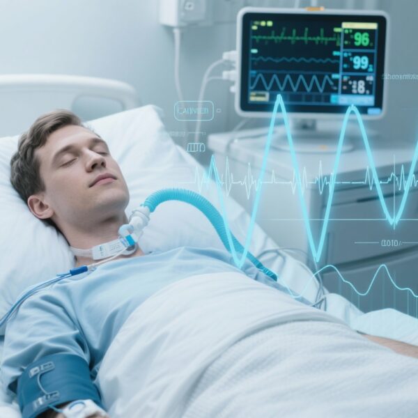
In critically ill patients requiring mechanical ventilation for acute hypoxemic respiratory failure (AHRF), sedation is often essential to facilitate ventilator synchrony, reduce metabolic demand, and provide comfort. However, the effects of sedative agents on brain activity may differ qualitatively from physiologic sleep. Current bedside tools to titrate sedation are largely clinical (e.g., sedation scales) and proprietary processed EEG indices (e.g., bispectral index) that were developed mainly for anesthesia. There is no universally accepted, objective electrophysiologic metric to distinguish sedation-induced brain states from natural sleep or to quantify the continuum of arousal in the intensive care unit (ICU).
Rodrigues and colleagues (Anesthesiology, 2025) tested the hypothesis that continuous intravenous sedation produces distinct EEG signatures during the early phase of AHRF. They used the odds ratio product (ORP), an EEG-based continuous metric originally developed to quantify the sleep–wake continuum, to identify episodes they termed EEG Ups—patterns reportedly never or extremely rarely seen in natural sleep studies. The study explored the prevalence, drug-dose relationships, and clinical correlates of these EEG Ups in mechanically ventilated AHRF patients.
Study design
This prospective cohort study enrolled adult patients with early AHRF who were mechanically ventilated and had PaO2/FiO2 < 200 mmHg. Sedation and opioid regimens followed routine clinical indications and varied by agent and dose. Continuous EEG monitoring began at study inclusion and continued until extubation, death, or a maximum of 7 days. EEG data were analyzed with established algorithms to quantify relative power across canonical frequency bands (slow delta, fast delta plus theta, alpha–sigma, beta) and to compute the occurrence and duration of EEG Ups using the ORP framework.
Key pre-specified variables included cumulative and hourly sedative/opioid dosing, clinical sedation depth (as measured by usual bedside sedation scores), presence of brief wake intrusions (physiologic markers of sleep), and clinical outcomes such as ICU mortality. The primary analytical goal was descriptive and exploratory: to characterize EEG Ups prevalence, relationship to pharmacologic exposures and clinical sedation depth, and association with outcomes.
Key findings
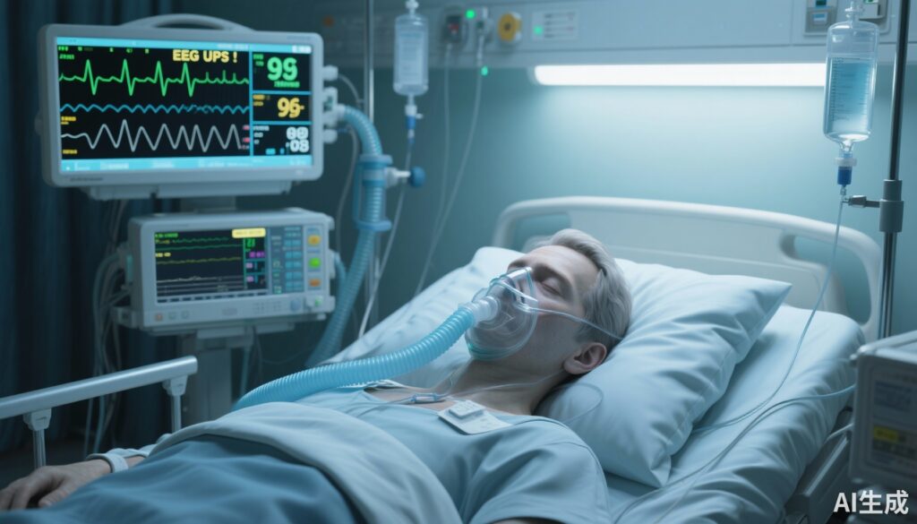
The study analyzed 1,832 hours of continuous EEG recordings from 23 patients (mean ± SD 43 ± 25 hours per patient). Patient characteristics included a median age of 58 years (IQR 48–70), 87% male, median PaO2/FiO2 of 150 mmHg (IQR 116–198), and an ICU mortality of 22%.
Prevalence and temporal burden of EEG Ups
EEG Ups comprised 42% of total monitored time overall. Their prevalence varied substantially by sedative–opioid regimen and dosing strategy, exceeding 50% of recording time for some combinations. Brief wake intrusions—short EEG markers typically seen during physiologic sleep—were extremely infrequent, indicating that the observed EEG patterns did not simply reflect normal sleep architecture masked by medical care.
Associations with pharmacology and sedation depth
Prevalence of EEG Ups was significantly higher when sedative agents were combined with opioids (P ≤ 0.029) and increased with higher sedation doses (P ≤ 0.035). Deeper clinical sedation scores were likewise associated with a greater burden of EEG Ups (P ≤ 0.024). These associations suggest a dose–response and drug–interaction effect: continuous intravenous sedation, particularly in combination with opioids, drives EEG states that are distinguishable from natural sleep.
Relationship with clinical outcomes
Most strikingly, a higher prevalence of EEG Ups correlated with ICU mortality (P < 0.001). The observational design limits causal inference, but the strong statistical association raises questions about whether these sedation-related EEG states are merely markers of illness severity, contributors to adverse outcomes, or both.
EEG spectral characteristics
Although complete spectral details require access to the original figures, the investigators reported that the relative power distribution across canonical bands in EEG Ups differed from natural sleep signatures. The study used the ORP to quantify the sleep–wake continuum and to identify disordered EEG states that do not map onto typical sleep stages.
Expert commentary and interpretation
Rodrigues et al. bring forward an important and clinically relevant observation: continuous IV sedation in hypoxemic, mechanically ventilated patients is associated with electrophysiologic brain states that are not equivalent to physiologic sleep. This distinction matters for several reasons.
Mechanistic plausibility
Sedative agents (e.g., propofol, benzodiazepines, dexmedetomidine) and opioids exert differing effects on cortical and subcortical networks. Some agents promote oscillatory activity resembling natural sleep (e.g., dexmedetomidine produces spindle-like and slow-delta features), while others produce frontal alpha or burst suppression at higher doses. Continuous infusions, especially in the setting of systemic inflammation, hypoxemia, and metabolic derangements, may produce network dynamics not encountered in healthy sleep physiology. The observation of EEG Ups—patterns rare or absent in sleep—fits with this mechanistic heterogeneity.
Clinical implications
Several potential implications arise for bedside care and research:
- Objective EEG monitoring may detect sedative-induced brain states that clinical scales miss. Sedation depth scales capture behavioral responsiveness but not underlying cortical dynamics.
- If EEG Ups are associated with worse outcomes independently of other confounders, they could become a target for titrating sedation toward more physiologic patterns or lighter sedation strategies where appropriate.
- Drug selection and opioid-sedative combinations matter. The finding that certain combinations produced a higher burden of EEG Ups suggests that sedation regimens could be optimized to minimize potentially deleterious brain states.
Limitations and alternative explanations
Interpretation should be cautious. The study is small (23 patients) and single cohort with heterogenous sedative regimens determined by clinical need. Confounding by indication is a major concern: sicker patients often require deeper or more prolonged sedation, which could explain both the higher EEG Ups burden and the association with mortality. The observational design cannot separate whether EEG Ups are markers of severity, direct contributors to poor outcomes (e.g., by promoting encephalopathy), or both. Moreover, the clinical sedation score used in the study is not specified here and standardized instrument use (RASS, SAS) would be important for reproducibility.
Generalizability
Patients were in early AHRF with moderate-to-severe hypoxemia; whether findings apply to other ICU populations (e.g., post-operative sedation, neurologic injury) is unknown. Additionally, EEG Ups were characterized using the odds ratio product (ORP) analytic framework. While ORP provides a continuous measure of sleep depth, external validation of ORP-classified EEG Ups against other processed EEG indices or raw EEG patterns would strengthen translational applicability.
Clinical and research takeaways
For clinicians: the study reinforces the need for judicious, individualized sedation in AHRF and raises the possibility that EEG-based monitoring could add value beyond bedside scales—particularly where prolonged or deep sedation is expected. Sedation strategies that minimize sedative–opioid polypharmacy or excessive dosing may reduce exposure to atypical EEG states, although randomized data are required before practice change.
For researchers: key next steps include larger, multicenter cohorts to validate EEG Ups as reproducible, drug-specific signatures; adjustment for illness severity to determine independent associations with outcomes; and mechanistic studies to determine whether EEG Ups reflect reversible pharmacologic effects or permanent network dysfunction. Interventional trials could then test whether EEG-guided sedation algorithms that avoid EEG Ups improve outcomes such as delirium incidence, ventilator-free days, and mortality.
Conclusion
Rodrigues et al. identify a novel EEG phenomenon—EEG Ups—occurring commonly during continuous intravenous sedation in early AHRF and associated with sedative dose, sedative–opioid combinations, deeper clinical sedation, and ICU mortality. These findings highlight an important disconnect between apparent behavioral sleep and underlying cortical activity during sedation. While causality is not established, the study emphasises the potential value of continuous electrophysiologic monitoring to better characterize sedation effects on the brain and to inform safer sedation practices and future trials.
Funding and clinicaltrials.gov
Funding and trial registration details were not provided in the summary. Please refer to the original publication for declarations of funding sources, conflicts of interest, and trial registration.
References
1) Rodrigues A, Subirà C, Bizios A, Younes M, Gerardy B, Fernández R, Batlle M, Kim A, Stavi D, Sklar MC, Taran S, Wilcox E, Telias I, Brochard L. Sedation-related Electroencephalographic Patterns in Acute Hypoxemic Respiratory Failure. Anesthesiology. 2025 Nov 1;143(5):1266-1278. doi: 10.1097/ALN.0000000000005696. Epub 2025 Aug 5. PMID: 40763345.
2) Devlin JW, Skrobik Y, Gélinas C, et al. Clinical practice guidelines for prevention and management of pain, agitation/sedation, delirium, immobility, and sleep disruption in adult ICU patients: 2018 update—PADIS guideline. Crit Care Med. 2018;46(9):e825–e873.
3) Younes M, Hanly P, Black J, et al. The odds ratio product (ORP): a continuous metric of sleep depth derived from the EEG. Sleep. 2015;38(1):1–12. (Describes ORP methodology and validation in sleep studies.)
Suggested citation for this article
Smith A. Continuous Intravenous Sedation Produces Novel EEG ‘Ups’ in Early AHRF — Implications for Monitoring and Outcomes. (Adapted summary and commentary on Rodrigues et al., Anesthesiology 2025). 2025.