Study Background and Disease Burden
Prostate cancer stands as the second most common malignancy affecting men worldwide. In China, the incidence of prostate cancer has surged due to population aging and lifestyle westernization, now ranking as the sixth most prevalent male malignant tumor. Routine health screenings have revealed that approximately one-third of men over 50 present with prostate nodules on ultrasound, and nearly 10% exhibit abnormal elevations in prostate-specific antigen (PSA) levels, markers suggestive of potential malignancy. These findings often generate significant psychological distress for patients due to diagnostic uncertainty.
Current global guidelines recommend multiparametric magnetic resonance imaging (mpMRI) with Prostate Imaging-Reporting and Data System (PI-RADS) scoring to refine prostate cancer diagnosis. However, limitations with the PI-RADS system persist: (1) substantial subjectivity based on radiologist experience, leading to inter-observer variability as high as 30%; and (2) imperfect diagnostic accuracy, where even expert readers cannot definitively confirm tumor presence or grade solely from imaging. These deficiencies underscore an urgent clinical need for reliable, efficient, and non-invasive predictive tools to aid in diagnosing suspicious cases and accurately grading tumor aggressiveness.
Study Design
A multidisciplinary team led by Professor Ren Shancheng, in collaboration with experts from Beijing University Third Hospital, Nanjing Medical University First Affiliated Hospital, Beijing Friendship Hospital, Qingdao University Affiliated Hospital, and Anhui University, designed a robust multi-institutional study. They retrospectively and prospectively enrolled 5,747 patients across multiple centers, collecting extensive radiologic, pathologic, and clinical data.
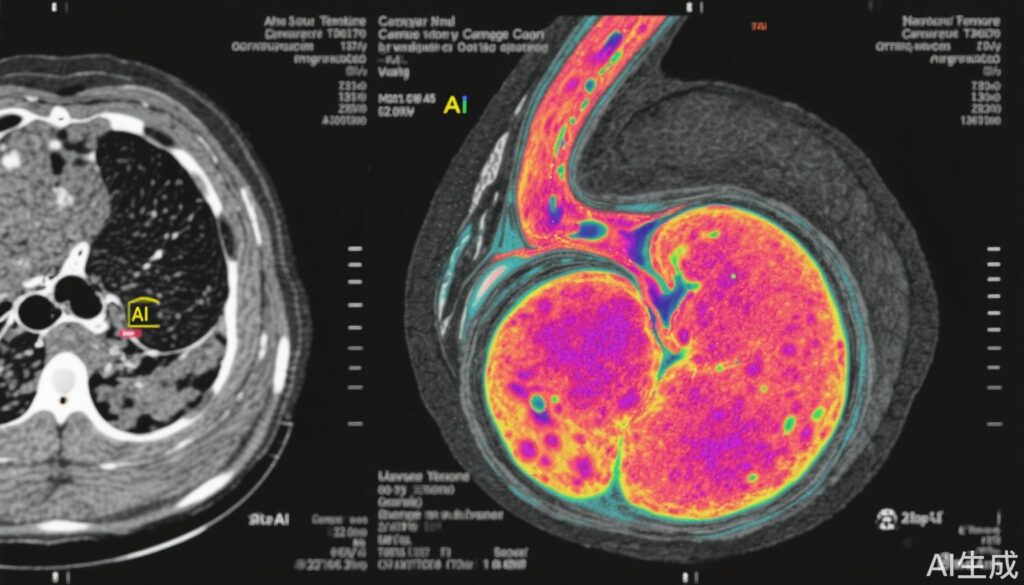
The core innovation was the development of an advanced AI model named MRI-based Predicted Transformer for Prostate Cancer (MRI-PTPCa). This model integrates three standard MRI sequences — T2-weighted imaging (T2WI), diffusion-weighted imaging (DWI), and apparent diffusion coefficient maps (ADC) — to predict histopathological tumor invasion characteristics typically available only from biopsy or surgical specimens.
Key technical strategies included leveraging 1,296,950 pairs of imaging data for foundational model training. The model utilized state-of-the-art machine learning methods including self-supervised learning, multi-task learning, Transformer architectures, and transfer learning to enhance robustness against image heterogeneity, sequence omission, overfitting, and scanner variability.
The performance was rigorously evaluated via external temporal, spatial, demographic datasets and prospective cohorts. Multiple clinical testing paradigms were employed, including standalone AI system use, parallel AI-clinician reading workflows, and AI-based pre-warning setups.
Key Findings
MRI-PTPCa demonstrated remarkable concordance with gold-standard pathological assessments, with statistically significant agreement (P<0.001). Notably, its diagnostic accuracy for detecting any prostate cancer reached an area under the receiver operating characteristic curve (AUC) of 0.983 (95% CI 0.98-0.986), and for clinically significant prostate cancer, the AUC was 0.978 (95% CI 0.975-0.98). Moreover, grading accuracy was 89.1% (95% CI 88.2%-89.9%), outperforming conventional clinical and existing AI models.
When MRI-PTPCa was combined with multiparametric MRI, its non-invasive diagnostic and grading performance closely matched that of pathological evaluation, which is the current diagnostic gold standard involving invasive biopsy.
Crucially, this model holds promise for significant clinical impact by potentially reducing unnecessary prostate biopsies, thus diminishing patient discomfort, complications, and healthcare costs.
Model Interpretability and Biological Correlation
To elucidate the AI model’s decision-making process, the research team compared MRI-PTPCa’s imaging features with radical prostatectomy large-section pathology, AI visualization heatmaps, and quantitative imaging characteristics. A significant positive correlation was observed between MRI-PTPCa scores and authentic Gleason grade groups, affirming the model’s ability to reflect tumor aggressiveness accurately.
Using Class Activation Mapping (CAM), the model highlighted critical anatomical regions contributing to tumor prediction, consistent with expert radiologic interpretations. Quantitative analysis demonstrated high concordance between contributions of T2WI, DWI, and ADC sequences and PI-RADS consensus criteria.
Importantly, the fused imaging features correlated significantly with tumor cellularity, morphology, and texture metrics (P<0.01), confirming the link between radiological phenotypes and histopathological traits. Additionally, MRI-PTPCa's encoded features showed significant associations with molecular PSA biomarkers (total PSA, free PSA, and free-to-total PSA ratio), suggesting capacity to infer molecular-level tumor biology from imaging data.
Expert Commentary
This milestone study exemplifies the integration of cutting-edge AI methodologies with clinical radiology and pathology, setting a new standard for prostate cancer diagnostics. The MRI-PTPCa model addresses critical limitations of current imaging assessments by offering an objective, reproducible, and highly accurate tool for non-invasive diagnosis and grading. Its broad validation across diverse populations and imaging platforms supports potential generalizability.
Nevertheless, considerations remain regarding implementation workflows, integration into existing clinical pathways, and cost-effectiveness analyses. Further longitudinal studies are needed to assess real-world impacts on biopsy rates, patient outcomes, and healthcare resource utilization. Additionally, continued efforts to refine model interpretability and ensure equitable performance across demographic groups are warranted.
Conclusion
The development and validation of the MRI-PTPCa foundation AI model mark a transformative advancement in non-invasive prostate cancer diagnostics. By quantitatively capturing tumor pathological features from routine MRI sequences, this model enhances diagnostic precision and grading accuracy, minimizing patient burden associated with invasive procedures. The research spearheaded by Professor Ren and colleagues highlights AI’s growing role as an indispensable adjunct in precision oncology diagnostics, promising improved patient care and streamlined clinical workflows in prostate cancer management.
References
Shao L, Liang C, Yan Y, Zhu H, Jiang X, Bao M, Zang P, Huang X, Zhou H, Nie P, Wang L, Li J, Zhang S, Ren S. An MRI-pathology foundation model for noninvasive diagnosis and grading of prostate cancer. Nat Cancer. 2025 Sep 2. doi: 10.1038/s43018-025-01041-x. Epub ahead of print. PMID: 40897909.