Highlight
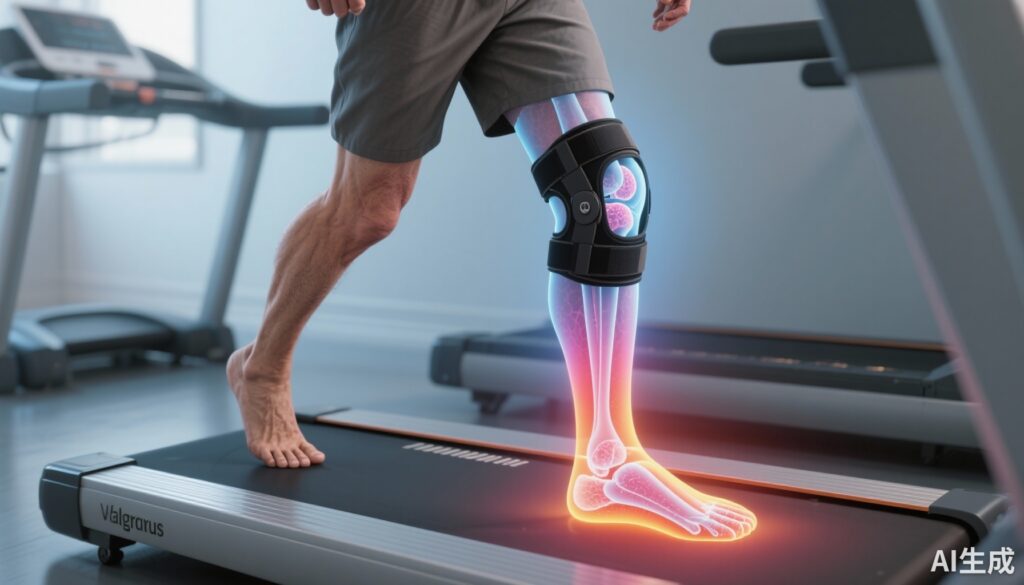
– Valgus bracing produced statistically significant reductions in medial tibial, medial femoral, and combined medial tibiofemoral cartilage contact pressures during specific phases of stance during walking in patients with varus‑malaligned medial knee osteoarthritis (OA).
– The largest observed reduction in maximum medial tibial cartilage pressure was 2.0 MPa (12.5% decrease) at 87% of stance.
– Bracing also shifted the center of pressure (CoP) laterally on the medial tibial and medial femoral cartilage early and mid‑stance, without detectable changes in patellofemoral cartilage pressure.
Background
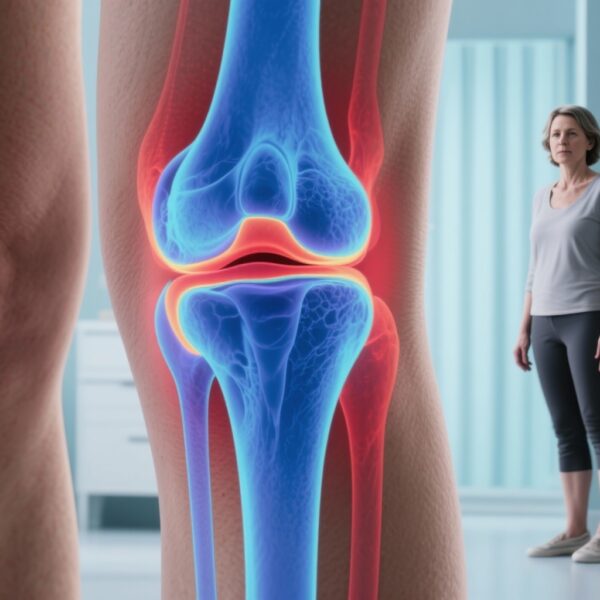
Medial compartment knee osteoarthritis commonly associates with varus malalignment, which increases the mechanical load borne by the medial tibiofemoral cartilage and is linked to pain and disease progression. Valgus knee bracing — intended to apply an external moment that counteracts varus alignment — is a conservative treatment option aimed at reducing medial compartment loading and improving symptoms.
Prior studies examining brace effects on whole‑joint kinetics (for example, medial compartment contact force or external knee adduction moment) have reported inconsistent or modest reductions. Tissue‑level mechanics — specifically cartilage contact pressure magnitude and the precise location of loading across cartilage surfaces — may better explain how bracing could reduce pain or slow structural damage. Starkey and colleagues (Clin Orthop Relat Res. 2025) used subject‑specific MRI geometry combined with an EMG‑informed musculoskeletal model and nonlinear elastic foundation contact simulations to estimate cartilage pressure during walking with and without valgus bracing in varus‑malaligned medial knee OA patients.
Study design
This secondary analysis used baseline data from a clinical trial cohort recruited in Melbourne, Australia (April–November 2019). Inclusion criteria included tibiofemoral radiographic OA, varus malalignment, age ≥ 50 years, and ≥ 3 months of knee pain. Of 28 participants originally enrolled, 25 had complete MRI data and were analyzed (mean ± SD age 64 ± 5 years; BMI 29.4 ± 3.1 kg/m2; 14 males, 11 females).
Each participant completed walking trials in unbraced and valgus‑braced conditions. A validated 12 degrees‑of‑freedom knee model with MRI‑derived cartilage morphology and ligament insertion points was combined with a calibrated, EMG‑informed neuromusculoskeletal model to simulate tissue contact mechanics. Tibiofemoral and patellofemoral cartilage contact simulations were performed across stance for four trials per condition using a nonlinear elastic foundation contact model.
Primary outcomes addressed two questions: (1) Did valgus bracing reduce estimated maximum and mean medial tibial, medial femoral, and patellar cartilage contact pressure during stance? (2) Did bracing change the center‑of‑pressure location on medial tibial, medial femoral, or patellar cartilage? Outcomes were expressed as MPa for pressures and as percentage of cartilage width/length (normalized) for CoP shifts. Time‑varying differences across stance were tested using statistical parametric mapping (one‑way repeated‑measures ANOVA); the largest pointwise differences across stance are reported with means and 95% CIs.
Key findings
Pressure magnitude
Valgus bracing reduced maximum medial tibial cartilage contact pressure at multiple portions of stance (7–15%, 26–40%, and 65–90% of stance). The largest between‑condition difference occurred at 87% of stance: unbraced mean ± SD 16.0 ± 4.3 MPa versus braced 14.0 ± 3.6 MPa (mean difference −2.0 MPa; 95% CI −3.5 to −0.6; p < 0.001), corresponding to a 12.5% reduction in peak maximum medial tibial pressure.
Comparable magnitude reductions were observed for mean tibial cartilage pressure and for maximum and mean medial femoral cartilage pressure during similar regions of stance. The patellofemoral cartilage contact pressure (both maximum and mean) did not differ between braced and unbraced walking across stance.
Location of loading (center of pressure)
On the medial tibial cartilage, braced walking shifted the center of pressure laterally relative to unbraced during early to mid‑stance (0–54% of stance) and more posteriorly in early stance (0–12% of stance). The largest lateral shift on medial tibia was at 4% stance (unbraced −0.8% ± 2.1%; braced 3.6% ± 3.8%; mean difference 4.4%; 95% CI 1.9% to 6.8%; p < 0.001). The largest posterior shift was at 5% stance (unbraced −5.0% ± 2.9%; braced −3.0% ± 3.5%; mean difference 2.0%; 95% CI 0.2% to 3.8%; p < 0.003).
On the medial femoral cartilage, the CoP was also shifted more laterally during braced walking, although no consistent anterior‑posterior CoP differences were seen. No significant CoP changes were observed on the patellar cartilage.
Clinical and statistical relevance
The observed peak reductions in medial tibiofemoral cartilage pressure were modest but consistent across participants and occurred during clinically relevant parts of stance (including late stance). The 12.5% peak reduction in maximum medial tibial pressure is physiologically plausible to modify cartilage mechanobiology or nociceptive drive, though a direct link to symptom improvement or structural modification remains to be established.
Expert commentary and interpretation
Starkey et al. expand our mechanistic understanding of how valgus bracing may function in varus‑aligned medial knee OA. Prior work focusing on whole‑joint metrics (for example, external knee adduction moment or estimated medial compartment contact force) has shown variable brace effects; the present study’s tissue‑level simulations indicate bracing can both reduce the magnitude of medial cartilage pressures and shift load laterally within the medial compartment.
These findings align with the mechanical rationale for valgus bracing: applying an external valgus moment reduces medial compartment loading and can alter contact mechanics. The lateral shift in CoP suggests a redistribution of load away from more medially concentrated contact regions, which could be relevant to pain if those regions correspond to areas of cartilage erosion, bone marrow lesions, or nociceptor‑rich subchondral bone.
However, several caveats temper interpretation. The study analyzed short‑term, immediate braced versus unbraced walking at baseline; it does not provide evidence that these mechanical changes persist during activities of daily living over time or that they translate into improvements in pain, function, or structural outcomes. Simulations depend on model assumptions (elastic foundation contact, material properties, cartilage thickness and cartilage/subchondral bone behavior) and on accurate EMG‑informed muscle force estimations; errors in these inputs can influence pressure estimates. The sample size was modest (n = 25), reflecting a secondary analysis, and participants represented a specific phenotype (older adults with radiographic medial TF OA and varus malalignment), which improves internal relevance but limits generalizability to broader OA populations.
Limitations
– Secondary analysis of baseline data with a modest sample size and potential selection bias. 3 of 28 initial participants were excluded due to incomplete MRI.
– Single activity (walking) and short‑term braced exposure; no assessment of longitudinal effects, brace adherence, or symptomatic outcomes linked to the mechanical changes.
– Modeling assumptions: elastic foundation contact models and constitutive cartilage properties are simplifications; tissue heterogeneity, depth‑dependent properties, and meniscal effects may not be fully captured.
– Brace specifics (type, fit, valgus moment magnitude) can vary in real world; the study does not report long‑term wear, comfort, or patient preference, which influence clinical uptake.
Clinical implications and next steps
For clinicians managing varus‑malaligned medial knee OA, this study provides biomechanically plausible evidence that properly applied valgus bracing can reduce medial tibiofemoral cartilage contact pressure and shift loading laterally during walking. These tissue‑level changes offer a mechanistic rationale to support selective use of valgus bracing in patients with symptomatic medial compartment OA and varus alignment, particularly when nonoperative strategies are sought.
Key unanswered questions for practice-oriented research include whether these immediate pressure reductions: (1) are maintained during long‑term brace use and across diverse activities; (2) correlate with clinically meaningful improvements in pain and function; and (3) reduce structural disease progression on imaging. Future randomized trials should integrate tissue‑level biomechanical endpoints with patient‑reported outcomes and structural imaging, and investigate which brace designs, fitting strategies, and patient phenotypes yield the greatest clinical benefit.
Conclusion
Starkey et al. provide subject‑specific simulation evidence that valgus bracing reduces medial tibiofemoral cartilage contact pressure and shifts loading laterally in varus‑malaligned medial knee OA during walking. These findings add mechanistic support to the use of valgus bracing but do not yet prove clinical effectiveness for pain relief or structural modification. Well‑designed prospective trials linking biomechanical changes to patient‑centred outcomes are needed to translate these results into treatment guidance and to refine brace design and patient selection.
Funding and clinicaltrials.gov
Funding and registration details are reported in Starkey SC et al., Clin Orthop Relat Res. 2025. Readers should consult the original article for full trial registration numbers, funders, and conflict of interest disclosures.
References
1. Starkey SC, Esrafilian A, Saxby DJ, Diamond LE, Korhonen RK, Hall M. Does Valgus Bracing Reduce Estimates of Medial Tibial, Medial Femoral, and Patella Cartilage Contact Pressure in Varus Malaligned Medial Knee Osteoarthritis? Clin Orthop Relat Res. 2025 Oct 1;483(10):1969-1982. doi: 10.1097/CORR.0000000000003611. PMID: 40668179; PMCID: PMC12453385.
2. Felson DT. Osteoarthritis as a disease of mechanics. Osteoarthritis Cartilage. 2013 Jan;21(1):10-5. doi: 10.1016/j.joca.2012.09.012. PMID: 23098545.


