Introduction
Holding a health checkup report and seeing the word “calcification” can make anyone’s heart skip a beat. It’s natural to worry about what this might mean for your health. However, calcification in organs is a common finding on medical imaging, often not a cause for alarm. Understanding the different types of calcification and their clinical significance can help reduce anxiety and guide appropriate medical follow-up.
The Science of Organ Calcification: Physiological vs. Pathological
Calcification refers to the accumulation of calcium salts in body tissues. These deposits show up frequently in medical imaging, such as X-rays and CT scans. Broadly, we classify calcification into two types:
– Physiological Calcification: This is a normal process essential for healthy development and maintenance of certain tissues. For example, calcium deposition is critical in bone and tooth formation. These calcium deposits are stable and part of normal biology.
– Pathological Calcification: This occurs when calcium salts deposit abnormally in tissues, usually due to injury, inflammation, or metabolic imbalance. Common causes include infections, chronic inflammation, tissue damage, and aging. Examples include scar tissue calcification after pneumonia, tuberculosis-related lung calcifications, and stones formed in the gallbladder or urinary tract. These deposits may affect organ function depending on their extent and location.
When Should Calcifications Raise Concerns?
Certain types of calcification warrant closer attention due to their potential as markers or contributors to disease. We will explore key organs commonly involved and provide guidance on when to follow up.
Lung Calcification
Lung calcifications often represent healed inflammation or infections such as old pneumonia or tuberculosis scars, similar to how scars appear on the skin. Occupational exposure to dusts like asbestos or coal can also lead to lung calcifications and in severe cases, pneumoconiosis (e.g., coal worker’s pneumoconiosis).
On CT scans, calcified lung nodules are typically benign. Patterns such as diffuse, central, layered, or “popcorn” calcifications indicate non-cancerous lesions. In contrast, non-calcified nodules require monitoring or biopsy based on size and density to rule out malignancy.
Breast Calcification
Breast calcifications are common findings on mammograms and have diverse causes, including glandular atrophy due to aging, benign proliferative changes, fat necrosis, and malignancies.
Benign calcifications typically appear coarse, round or ring-shaped. Suspicious calcifications display segmental, clustered, or fine linear branching patterns. Radiologists use the BI-RADS (Breast Imaging Reporting and Data System) classification to guide management:
| BI-RADS Category | Meaning | Recommendation |
|——————-|———|—————-|
| 0 | Incomplete; further imaging needed | Additional imaging required |
| 1–3 | Probably benign | Routine follow-up |
| 4 | Suspicious abnormality | Biopsy suggested |
| 5 | Highly suggestive of malignancy | Biopsy necessary |
Thyroid Calcification
Calcifications in the thyroid gland occur in both benign and malignant conditions. They are categorized into microcalcifications, coarse calcifications, and peripheral calcifications. Microcalcifications are especially notable as an important warning sign for papillary thyroid carcinoma.
Ultrasound evaluation using TI-RADS (Thyroid Imaging Reporting and Data System) assists clinicians in determining malignancy risk and necessary interventions, including biopsy or surveillance.
Prostate Calcification
Prostate calcifications result commonly from chronic prostatitis or benign prostatic hyperplasia, leading to obstruction and calcified secretions. Most prostate calcifications are asymptomatic. However, those related to chronic inflammation may accompany pelvic pain.
In the absence of urinary symptoms such as urgency, frequency, or difficulty, special treatment is generally unnecessary—routine prostate ultrasound monitoring suffices.
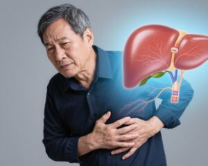
Liver Calcification
Inflammation from infections or parasites frequently causes liver calcification. Both benign tumors like hemangiomas and malignant lesions such as hepatocellular carcinoma can present with calcification.
Most liver calcifications detected incidentally are benign; however, their size, distribution, and correlation with clinical findings determine the management strategy, which often includes periodic imaging follow-up.
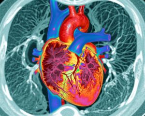
Coronary Artery Calcification
Coronary artery calcification (CAC) increases with aging and promotes vessel stiffening, impairing coronary blood flow. Risk factors include dyslipidemia, diabetes, hyperparathyroidism, chronic kidney disease, and hypercalcemia.
CAC can be quantitatively assessed using coronary CT angiography or invasive coronary angiography to identify stenosis risk. Identification mandates stringent management of underlying conditions, lifestyle modification, and regular cardiovascular monitoring.
Common Misconceptions About Calcification
| Misconception | Reality |
|—|—|
| Calcification always means cancer | Most calcifications are benign, especially when in typical patterns or locations.
| All calcifications require immediate surgery or biopsy | Many calcifications are monitored over time unless suspicious features arise.
| Calcification is solely caused by high calcium intake | Calcification is often secondary to tissue injury, inflammation, or metabolic disease rather than diet alone.
Practical Advice on Coping with Calcification Findings
– Stay calm and consult your healthcare provider to interpret the imaging findings in clinical context.
– Adhere to follow-up imaging schedules as recommended.
– Maintain a heart-healthy and anti-inflammatory lifestyle: balanced diet, regular exercise, and smoking cessation.
– Manage underlying chronic conditions such as diabetes, hypertension, and lipid disorders.
– For suspicious calcifications, proceed with recommended diagnostic tests like biopsies promptly.
Expert Recommendations
Dr. Emily Chen, a radiologist specializing in thoracic imaging, emphasizes: “Calcifications are commonly benign but must be viewed alongside patient history and other imaging features. Multidisciplinary evaluation ensures timely and accurate diagnosis.”
Dr. Michael Thompson, an endocrinologist, notes: “Thyroid and breast calcifications require careful ultrasound evaluation. The TI-RADS and BI-RADS systems standardize reporting and help guide appropriate interventions without over-treatment.”
Patient Scenario: John’s Unexpected Chest CT Result
John, a 58-year-old man with a history of smoking but otherwise healthy, underwent a routine health checkup including a chest CT scan. The report noted several calcified nodules in his lungs.
Initially anxious, John consulted his pulmonologist who explained that these calcifications were likely residual scars from past infections, common in people with certain occupational exposures. There was no sign of active disease or malignancy. John was advised to continue regular health checks and avoid further lung irritants.
This case illustrates the importance of understanding the nature of calcifications and the value of expert guidance to avoid unnecessary panic.
Conclusion
Calcifications detected during routine health screenings are common and often benign. Differentiating physiological from pathological calcifications and recognizing patterns suggestive of malignancy or serious disease is crucial.
Understanding the specific organ context, risk factors, and imaging features guides appropriate management ranging from simple monitoring to biopsy or intervention.
Staying informed, consulting healthcare professionals, and following recommended follow-up are key to navigating these findings safely and confidently.
References
1. Johnson KM, et al. Coronary artery calcium scoring: evidence and recommendations for clinical use. Cleve Clin J Med. 2020;87(9):571-582.
2. Kim SH, et al. Characterization of Thyroid Nodules and Microcalcifications: The Role of Ultrasound and FNAB. Diagnostics. 2021;11(12):2375.
3. Bassett LW, et al. BI-RADS® Atlas: Breast Imaging Reporting and Data System. American College of Radiology; 2013.
4. Gossmann A, et al. Lung Calcification—Incidental Findings and Clinical Impact. Respir Med. 2019;158:58-64.
5. Feng JC, et al. Prostate calcifications in men with chronic prostatitis/chronic pelvic pain syndrome. Asian J Androl. 2015;17(2):257-259.