Highlights
– New human-based evidence identifies impaired mitochondrial calcium uptake as a central, previously underappreciated mechanism in atrial fibrillation (AF).
– Subcellular structural remodeling — increased sarcoplasmic reticulum (SR)–mitochondria distance and microtubule destabilization — disconnects Ca2+ supply from mitochondrial ATP production, promoting energetic failure and oxidative stress.
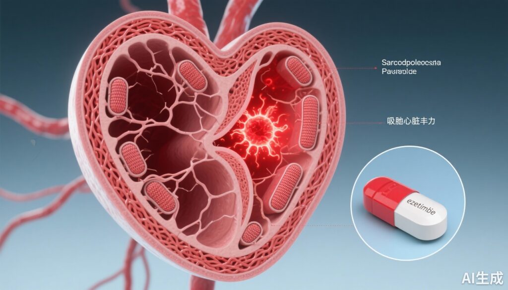
– In human atrial myocytes and hiPSC-derived cardiomyocytes, targeted antioxidant MitoTEMPO partially rescues dysfunction; the cholesterol absorption inhibitor ezetimibe unexpectedly enhances mitochondrial Ca2+ uptake, stabilizes electrophysiology, and is associated with lower AF burden in a retrospective cohort.
Background and clinical context
Atrial fibrillation (AF) is the most common sustained cardiac arrhythmia and a major contributor to stroke, heart failure, and health-care utilization worldwide. Current AF management focuses on anticoagulation to prevent thromboembolism, rate and rhythm control using drugs or ablation, and modification of risk factors. Nevertheless, recurrence rates after rhythm-control strategies remain high and antiarrhythmic drugs have limited efficacy and nontrivial toxicity. A deeper mechanistic understanding of AF pathogenesis at the cellular and subcellular level is needed to develop interventions that target upstream drivers rather than only surface membrane ion channels or triggers.
Study design and experimental approach
The study by Pronto et al. (Circ Res. 2025) combined human tissue analysis, cellular physiology, advanced imaging, and translational pharmacology to interrogate mitochondrial calcium handling in AF. Key elements included:
- Ex vivo analysis of right atrial appendage tissue obtained from patients undergoing cardiac surgery, with separation of atrial myocytes for functional assays.
- High-resolution live-cell measurements of cytosolic and mitochondrial Ca2+ dynamics under basal conditions and β-adrenergic stimulation (isoproterenol) to simulate increased workload.
- Biochemical assays of mitochondrial redox cofactors (NADH, FADH2) and reactive oxygen species (ROS) production.
- Electron tomography and super-resolution microscopy to quantify nano-scale organization of SR–mitochondria contacts and microtubule architecture.
- Mechanistic recapitulation using human induced pluripotent stem cell–derived cardiomyocytes (hiPSC-CMs) with pharmacologic or cytoskeletal perturbation to model microtubule instability.
- Intervention studies in vitro using the mitochondrial-targeted antioxidant MitoTEMPO and the cholesterol absorption inhibitor ezetimibe to test rescue of mitochondrial Ca2+ uptake and electrophysiologic stability.
- A retrospective cohort analysis evaluating AF burden among patients with paroxysmal AF who were receiving ezetimibe versus those who were not (methodological and covariate details reported in the original paper).
Key findings
The central observations link disrupted mitochondrial Ca2+ handling to energetic failure, oxidative stress, and arrhythmogenic calcium leak in AF:
1) Impaired mitochondrial Ca2+ uptake under physiologic stimulation
In atrial myocytes from patients with AF, mitochondrial Ca2+ transients in response to workload increase or β-adrenergic activation were markedly reduced compared with non-AF controls. This indicates that during tachyarrhythmic stress, mitochondria in AF fail to receive the cytosolic Ca2+ signals that normally stimulate the tricarboxylic acid (TCA) cycle and oxidative phosphorylation to match ATP supply to demand.
2) Bioenergetic compromise and increased oxidative stress
Concomitant with reduced mitochondrial Ca2+ uptake, measures of mitochondrial redox state showed impaired NADH/FADH2 regeneration, consistent with diminished substrate provision to the electron transport chain. Mitochondrial antioxidant capacity was decreased and ROS levels were elevated in AF myocytes, linking defective Ca2+ signaling to oxidative injury.
3) Subcellular structural remodeling — the SR–mitochondria “microdomain” is disrupted
Electron tomography and super-resolution imaging demonstrated increased nanoscale distance between SR and mitochondria in AF atrial tissue and disordered spatial arrangement. Microtubule networks that help position organelles were destabilized. The authors analogized this to a breakdown of a high-efficiency “calcium delivery highway”, resulting in failed Ca2+ transfer to mitochondria despite preserved cytosolic Ca2+ transients.
4) Microtubule disruption is sufficient to recapitulate the phenotype
Deliberate perturbation of microtubules in hiPSC-CMs reproduced reduced mitochondrial Ca2+ uptake and increased spontaneous Ca2+ release events (SCaEs), electrophysiologic abnormalities that can act as focal triggers for AF. These results support a causal role for cytoskeletal instability and organelle mispositioning in mitochondrial dysfunction.
5) Antioxidant rescue implicates ROS in a self‑reinforcing loop
Application of the mitochondrial-targeted antioxidant MitoTEMPO partially restored mitochondrial Ca2+ uptake and reduced abnormal Ca2+ release, indicating that oxidative stress is an important mediator that both follows and amplifies mitochondrial dysfunction.
6) Ezetimibe improves mitochondrial Ca2+ handling and is associated with lower AF burden
Surprisingly, ezetimibe augmented mitochondrial Ca2+ transient amplitude in AF atrial myocytes, prolonged action potential duration (suggesting greater electrical stability in this model), and reduced SCaEs. In a retrospective clinical cohort analysis included in the study, patients with paroxysmal AF who were on ezetimibe had a lower AF burden compared with matched controls. The authors emphasize these findings are exploratory and hypothesis-generating, but they raise the possibility that the drug’s effect may derive from restoring subcellular coupling and reducing oxidative stress rather than direct ion channel blockade.
Biological and mechanistic interpretation
The data present a coherent mechanistic model: during AF, excessive atrial activation increases energetic demand. Normal excitation–contraction coupling uses local cytosolic Ca2+ microdomains to activate mitochondrial dehydrogenases and boost ATP production. When SR–mitochondria contacts are lost and microtubules destabilize, mitochondrial Ca2+ uptake is impaired; mitochondrial dehydrogenase activation and NADH/FADH2 supply to the respiratory chain fall, ATP production becomes inadequate, and ROS accumulates. ROS further damages microtubules and calcium-handling proteins (including ryanodine receptors), promoting spontaneous Ca2+ release and electrical instability — a vicious cycle that sustains AF.
The observation that MitoTEMPO and ezetimibe interrupt parts of this cycle supports both redox modulation and organelle targeting as promising therapeutic strategies. While the molecular target by which ezetimibe influences mitochondrial Ca2+ handling remains unknown, potential mechanisms include modification of membrane lipid composition, effects on intracellular cholesterol trafficking, or off-target interactions that stabilize organelle contacts or attenuate oxidative stress.
Expert commentary and limitations
Strengths of the study include the use of human atrial tissue, multi-modal imaging, functional assays at both the organelle and cellular level, and translational pharmacologic tests. These features make the mechanistic conclusions particularly compelling for human AF.
However, important limitations temper immediate clinical translation:
- Sampling was from right atrial appendages of surgical patients — AF is a heterogeneous disease and left atrial-specific remodeling may differ.
- The retrospective clinical analysis of ezetimibe and AF burden is hypothesis-generating and subject to confounding by indication, concomitant statin use, and differences in comorbidities or health-care behavior. It cannot prove causality.
- Detailed molecular targets and signaling pathways linking ezetimibe to mitochondrial Ca2+ uptake were not defined; whether effects are direct on cardiomyocytes or mediated via systemic metabolic changes remains to be established.
- Preclinical rescue with MitoTEMPO is encouraging but does not substitute for well-designed clinical trials evaluating arrhythmic endpoints and long-term safety of targeting mitochondrial redox state in humans.
Given these caveats, the findings are best viewed as a conceptual advance: AF is not merely a membrane-ion-channel disease but also an organelle and energy-handling disorder. Interventions that restore SR–mitochondria coupling, stabilize cytoskeletal architecture, or reduce mitochondrial oxidative stress merit further investigation.
Translational implications and next steps
The study suggests several near- and mid-term research priorities:
- Prospective observational studies with careful adjustment and mechanistic biomarker collection (e.g., circulating oxidative stress markers, myocardial imaging of energetics) to further examine the association between ezetimibe and AF outcomes.
- Randomized controlled trials (RCTs) in selected AF populations (for example, patients with frequent paroxysms and evidence of metabolic/oxidative stress) testing ezetimibe versus placebo on AF recurrence, burden (by continuous monitoring), and safety. Trials should prespecify mechanistic substudies using atrial tissue (where ethically available) or advanced imaging.
- Further mechanistic laboratory work to identify molecular targets: does ezetimibe affect mitochondrial Ca2+ uniporter (MCU) complex function, mitochondrial-associated membrane (MAM) proteins that physically tether SR and mitochondria, or cytoskeletal stabilizers?
- Development and testing of targeted organelle-protective therapies — selective stabilizers of SR–mitochondria contacts, microtubule-protective agents with cardiac safety, and mitochondrial-specific antioxidants — in preclinical AF models and, subsequently, in carefully designed human trials.
Conclusion
Pronto et al. provide compelling human evidence that impaired mitochondrial Ca2+ handling — rooted in subcellular structural remodeling and amplified by oxidative stress — is central to AF pathogenesis. The unexpected signal that the widely used lipid-lowering agent ezetimibe can restore mitochondrial Ca2+ signaling and reduce arrhythmogenic Ca2+ release in vitro, together with an associated lower AF burden in exploratory clinical analyses, opens a new paradigm: targeting energy supply, organelle coupling, and oxidative stress to prevent or treat AF. These data justify mechanistic investigations and prospective clinical trials but should not yet change clinical practice.
Funding and clinicaltrials.gov
Funding sources are reported in the original publication (Pronto et al., Circ Res. 2025). There is no registered randomized clinical trial testing ezetimibe for AF prevention reported within this paper; prospective registration and trial design will be required prior to clinical implementation.
References
1. Pronto JRD, Mason FE, Rog‑Zielinska EA, et al. Impaired Atrial Mitochondrial Calcium Handling in Patients With Atrial Fibrillation. Circ Res. 2025 Oct 15. doi: 10.1161/CIRCRESAHA.124.325658. Epub ahead of print. PMID: 41090220.
2. Cannon CP, Blazing MA, Giugliano RP, et al.; IMPROVE‑IT Investigators. Ezetimibe Added to Statin Therapy after Acute Coronary Syndromes. N Engl J Med. 2015;372(25):2387–2397. doi:10.1056/NEJMoa1410489.


