Highlight
- The AI platform Diagnocat showed high sensitivity (up to 93.9%) in detecting periapical radiolucencies (PARLs) in CBCT scans of non-root-filled molars.
- Specificity was moderate, indicating potential for overdiagnosis, particularly in postoperative scans.
- Post-root canal treatment scans demonstrated significantly reduced positive predictive value (PPV) and F1 scores, suggesting diagnostic challenges post-treatment.
- The findings emphasize the complementary role of AI tools with expert human evaluation in endodontic diagnosis.
Study Background and Disease Burden
Periapical periodontitis, a common inflammatory lesion at the apex of a tooth root, often results from pulpal infection. Accurate detection of periapical radiolucencies (PARLs) is critical for diagnosis, treatment planning, and prognosis in endodontics. Traditional radiographic modalities such as periapical and panoramic radiographs have limitations in sensitivity and specificity, especially in complex anatomical regions like molars.
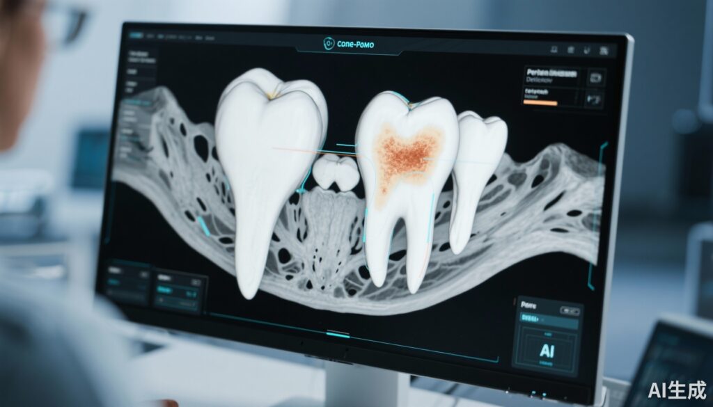
Cone-beam computed tomography (CBCT) offers superior spatial resolution and three-dimensional visualization, improving diagnostic confidence. However, interpreting CBCT scans requires expertise and can be time-consuming. Artificial intelligence (AI)-based tools promise to enhance diagnostic efficiency by automating lesion detection. The Diagnocat platform represents a convolutional neural network-based AI tool designed to detect PARLs on CBCT images.
Despite increasing integration of AI in dentistry, robust clinical validation of such platforms is necessary to ensure accuracy and reliability before widespread adoption. This study therefore aims to rigorously evaluate the diagnostic performance of Diagnocat in molars, comparing its effectiveness in untreated non-root-filled teeth as well as after root canal treatment.
Study Design
This retrospective study analyzed a dataset of cone-beam computed tomography scans from 134 molars comprising 327 roots, stratified by treatment status into preoperative (non-root-filled) and postoperative (root-filled) groups.
The Diagnocat AI platform was applied to these CBCT images to detect the presence of periapical radiolucencies. The AI outputs were compared against the reference standard established independently by two experienced endodontists, who reviewed the scans for PARLs without access to the AI results.
Diagnostic accuracy metrics were calculated for both tooth-level and root-level assessments. Evaluated metrics included sensitivity, specificity, accuracy, positive predictive value (PPV), negative predictive value (NPV), F1 score (harmonic mean of precision and recall), and area under the receiver operating characteristic curve (AUC-ROC).
Key Findings
In non-root-filled molars (preoperative scans), Diagnocat demonstrated:
– High sensitivity: 93.9% at tooth level and 86.2% at root level, indicating strong capability to detect true lesions.
– Moderate specificity: 65.2% (teeth) and 79.9% (roots), suggesting a tendency toward some false positives.
– Accuracy was 79.1% (teeth) and 82.6% (roots), reflective of balanced performance.
– PPV ranged from 71.8% to 75.8%, and NPV was notably high at 91.8% to 88.8%, illustrating reliable negative assessments.
– F1 scores were 81.3% (teeth) and 80.7% (roots), denoting overall good prediction quality.
– AUC-ROC values were 0.76 (teeth) and 0.79 (roots), indicative of moderate diagnostic efficacy.
Conversely, postoperative scans of root-filled molars showed considerably poorer performance:
– PPV dropped to 54.2% at the tooth level and 46.9% at the root level.
– F1 scores declined to 67.2% (teeth) and 59.2% (roots).
These findings suggest AI’s diminished reliability in detecting PARLs post-treatment, possibly due to artifacts, altered radiographic features from filling materials, or healing changes complicating pattern recognition.
Expert Commentary
The use of AI in dental radiology is an evolving frontier. Diagnocat’s high sensitivity in untreated molars is clinically valuable for initial diagnosis, reducing missed lesions. However, moderate specificity calls for caution as false positives may precipitate unnecessary interventions or anxiety. This underscores the crucial role of clinician oversight to contextualize AI-derived findings within comprehensive clinical evaluation.
The observed reduction of accuracy in postoperative scans highlights a key limitation of current AI models: difficulty distinguishing complex post-therapeutic changes from pathological lesions. Enhanced training datasets including diverse postoperative images, advanced algorithms compensating for beam hardening artifacts, or integrating clinical data might improve future performance.
From a biological standpoint, periapical radiolucencies represent inflammatory bone resorption, appearing as hypodense areas on CBCT. Changes induced by root filling materials can mimic or mask such findings, necessitating interpretative skill beyond raw image analysis.
Conclusion
Diagnocat demonstrates promising diagnostic accuracy in detecting periapical radiolucencies on CBCT scans of non-root-filled molars, characterized by high sensitivity and reasonable overall accuracy. However, its moderate specificity warrants that AI diagnosis be considered an adjunct to, not a replacement for, expert clinical judgment to mitigate overdiagnosis risks.
Diagnostic performance notably deteriorates in postoperative root-filled teeth, limiting Diagnocat’s current utility for disease monitoring after root canal therapy. Continued refinement of AI algorithms, validation in larger clinical cohorts, and integration of multimodal data are essential to enhance reliability across all clinical scenarios.
Clinicians should remain actively engaged in interpreting AI-generated findings and employ these tools to augment, not substitute, their diagnostic process. Future research should explore training AI systems on comprehensive postoperative datasets and assessing impact on patient outcomes.
References
Allihaibi M, Koller G, Mannocci F. Diagnostic accuracy of an artificial intelligence-based platform in detecting periapical radiolucencies on cone-beam computed tomography scans of molars. J Dent. 2025 Sep;160:105854. doi: 10.1016/j.jdent.2025.105854. Epub 2025 May 31. PMID: 40456383.
Patel S, Dawood A, Ford TP. The potential applications of cone beam computed tomography in the management of endodontic problems. Int Endod J. 2007;40(10):818-30.
Schwendicke F, Golla T, Dreher M. Artificial intelligence in dental diagnostics—state of the art and future perspectives. Br Dent J. 2021;230(8):477-485.
Setzer FC, Shah S, Kohli MR, Karabucak B, Kim S. Cone-beam computed tomography in endodontics — A review. J Endod. 2012;38(9):1338-1345.


