Background and Disease Burden
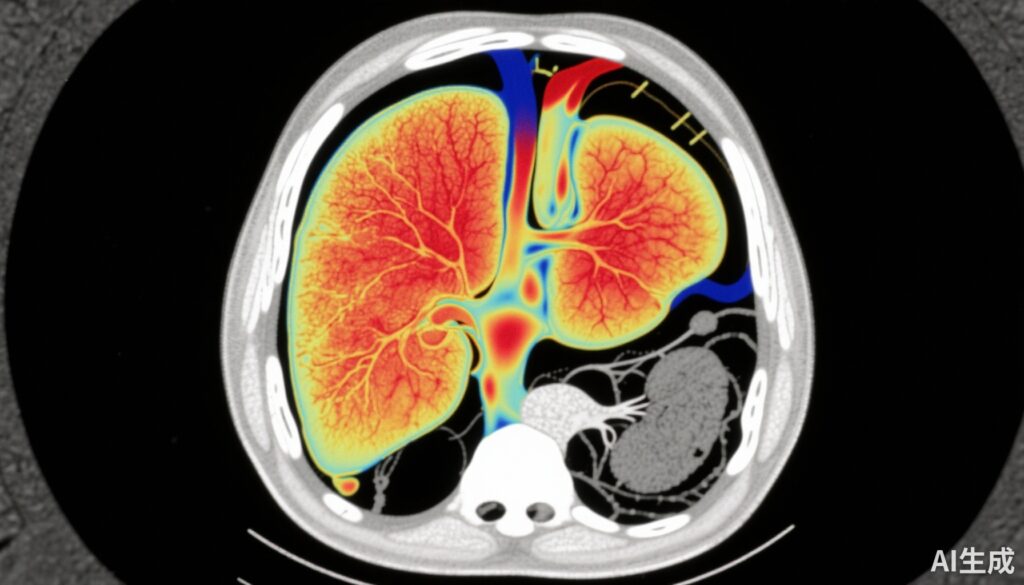
Chronic liver disease (CLD) presents a significant global health burden due to its progressive nature that can culminate in cirrhosis, liver failure, and hepatocellular carcinoma. Early identification of patients at risk of disease progression remains paramount to guide appropriate monitoring and therapeutic interventions. Traditional liver biopsy, regarded as the reference standard for fibrosis assessment, is limited by invasiveness, sampling variability, and patient reluctance. Consequently, noninvasive imaging tests (NITs) have increasingly become integrated into clinical care pathways, facilitating safer and potentially more comprehensive liver evaluation. Magnetic resonance (MR)-based techniques, specifically multiparametric MRI (mpMRI) including corrected T1 (cT1) and magnetic resonance elastography (MRE), offer quantitative assessments of liver fibrosis, inflammation, fat, and iron content. However, real-world data regarding their applicability and prognostic accuracy across diverse liver disease etiologies remain sparse. This study aimed to evaluate clinical use patterns, referral trends, and the prognostic value of cT1 and MRE in predicting liver disease progression in a heterogeneous patient cohort under routine clinical conditions, with a particular focus on steatotic liver disease (SLD), a growing public health challenge associated with obesity and metabolic syndrome.
Study Design and Methods
This prospective observational study enrolled 256 patients referred for abdominal MR imaging over an 18-month period irrespective of liver disease etiology or referral pathway, reflecting real-world clinical practice. Baseline demographics included a mean age of 53 years, balanced gender distribution (51% female), and a substantial prevalence of obesity (48% with BMI > 30 kg/m2).
Liver fibrosis was quantified using MRE, with hepatic stiffness measurements expressed in kilopascals (kPa). Disease severity was assessed by mpMRI parameters including iron-corrected T1 (cT1) reflecting fibroinflammatory activity, liver fat content (LFC), and liver iron concentration. Comparative serologic fibrosis risk was gauged by FIB-4 scores, though the focus remained on imaging modalities. Statistical analyses comprised t-tests for subgroup comparisons, Kaplan-Meier survival analyses for longitudinal progression outcomes, and receiver operating characteristic (ROC) curve analysis to compare predictive performance (area under the curve, AUC). The primary endpoint was liver disease progression defined by clinical, laboratory, or imaging criteria during follow-up.
Key Findings
A significant majority (66%) of the cohort had steatotic liver disease (SLD), consistent with the obesity prevalence. Several important observations emerged:
– Among patients with low MRE values ( 875 ms), signifying ongoing fibroinflammatory activity despite low stiffness.
– Elevated cT1 was consistently observed in patients with high MRE (> 5 kPa), confirming that active disease tends to coexist with advanced fibrosis.
– During longitudinal follow-up, those with low fibrosis risk by MRE but elevated disease activity by cT1 had a significantly greater risk of clinical disease progression (hazard ratio [HR] 3.1, p = 0.0035), highlighting cT1 as an independent prognostic marker beyond fibrosis.
– In the subgroup of SLD patients, cT1 showed superior discriminatory capability for predicting progression (AUC 0.71) compared to liver fat content (AUC 0.64) and MRE (AUC 0.53), underscoring its clinical importance in fatty liver disease where fibrosis may be less advanced but inflammation drives progression.
These results emphasize the complementary nature of MRE and cT1, where reliance on fibrosis measurement alone may underestimate risk, particularly in early or inflammatory disease stages.
Expert Commentary and Clinical Implications
This real-world assessment validates growing evidence that multiparametric MRI provides critical insights into chronic liver disease beyond fibrosis quantification. The intriguing finding that elevated cT1 identifies patients at high risk despite reassuring MRE values suggests that fibroinflammatory processes may precede or occur independently of fibrotic remodeling detectable by stiffness measurements alone. Recognizing this fibroinflammatory activity can prompt earlier therapeutic interventions, such as lifestyle modification or pharmacotherapy, potentially arresting progression before irreversible fibrosis ensues.
Clinicians should consider integrating both MRE and cT1 into routine liver disease assessment algorithms, especially for SLD populations, where standard fibrosis markers are limited in prognostic accuracy. These multiparametric markers support personalized risk stratification and may guide tailored surveillance intervals and treatment decisions.
However, limitations exist, including the single-center observational design and the lack of direct biopsy correlation for validation. Moreover, generalizability to less specialized centers or broader populations requires further study. Future research should explore how combining imaging biomarkers with clinical and serological data can refine prognostication and optimize management pathways.
Conclusions
This study reinforces the prognostic value of liver corrected T1 imaging alongside magnetic resonance elastography in chronic liver disease management. Patients exhibiting low fibrosis risk via MRE but elevated cT1 constitute a high-risk group with a threefold increase in progression likelihood. Particularly in steatotic liver disease, cT1 outperforms traditional fat and stiffness measurements in predicting disease worsening. Integrating both fibrotic and inflammatory imaging biomarkers into standard clinical protocols offers a more nuanced and predictive evaluation of liver disease, facilitating earlier interventions and improved patient outcomes.
Funding and Disclosures
The referenced study was published without specific funding disclosures noted. No clinical trial registration number was provided.
References
Corey KE, Nakrour N, Bethea ED, Shay JE, Andersson KL, Bhan I, Friedman LS, Kambadakone AR, Dichtel LE, Chung RT, Harisinghani M. Real-World Assessment of Liver Corrected T1 and Magnetic Resonance Elastography in Predicting Liver Disease Progression. Liver Int. 2025 Sep;45(9):e70280. doi: 10.1111/liv.70280. PMID: 40810289; PMCID: PMC12351529.
Younossi ZM, Golabi P, de Avila L, Paik JM, Srishord M, Fukui N, Henry L, Younossi I, Racila A, Afendy A. The Global Epidemiology of NAFLD and NASH in Patients with Type 2 Diabetes: Current Evidence and Perspectives. Diabetes Metab Syndr Obes. 2020;13:1313-1323.
Allen AM, Van Houten HK, Sangaralingham LR, Larson JJ, Therneau TM, Shah ND, Doucette JT, Wieland AL, Kamath PS, Sanderson SO. Fibrosis Progression in Nonalcoholic Fatty Liver vs Nonalcoholic Steatohepatitis: A Retrospective Cohort Study. Hepatology. 2018 Jul;68(1):104-115.
Banerjee R, Pavlides M, Tunnicliffe EM, Piechnik SK, Sarania N, Philips R, Collier JD, Booth JC, Schneider JE, Wang LM, et al. Multiparametric Magnetic Resonance for the Non-invasive Diagnosis of Liver Disease. J Hepatol. 2014;60(1):69-77.
Abbreviations
SLD: Steatotic Liver Disease
CLD: Chronic Liver Disease
mpMRI: Multiparametric Magnetic Resonance Imaging
MRE: Magnetic Resonance Elastography
cT1: Corrected T1
LFC: Liver Fat Content
HR: Hazard Ratio
AUC: Area Under the Curve
BMI: Body Mass Index


