Highlight
- AI assistance significantly reduces false positive diagnoses in detecting periapical radiolucencies (PRs) on panoramic radiographs.
- Junior dentists experience the greatest improvements in diagnostic accuracy and confidence with AI support.
- AI guidance shifts treatment decisions toward more conservative management, potentially reducing overtreatment.
Study Background and Disease Burden
Periapical radiolucencies (PRs) are radiographic manifestations of pathological processes commonly associated with pulp necrosis and apical periodontitis, conditions that are prevalent and clinically significant in dental practice. Detecting PRs accurately is essential to guide appropriate endodontic or restorative treatment, avoid unnecessary interventions, and ensure optimal patient outcomes. Panoramic radiography is widely used owing to its broad coverage and cost-effectiveness, but its sensitivity and specificity for detecting PRs are limited compared to cone-beam computed tomography (CBCT). Interpretation variability among dentists, notably across different experience levels, further compounds diagnostic inconsistency and may lead to over- or undertreatment.
Recent advances in artificial intelligence (AI) and machine learning have introduced promising tools for image analysis and diagnostic support. Incorporating AI into panoramic radiograph interpretation could standardize assessments, enhance diagnostic accuracy, and improve treatment planning. However, clinical evidence from rigorous trials evaluating AI’s impact on dental diagnostic practice remains sparse. This randomized controlled trial was designed to address this gap by evaluating AI assistance’s effects on diagnostic accuracy, confidence, and treatment decisions when assessing PRs on panoramic images.
Study Design
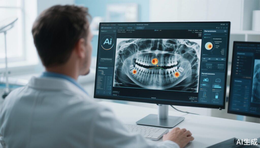
This randomized, cross-over controlled trial involved 30 dentists with varying clinical experience levels — including junior practitioners and experienced clinicians. Each participant evaluated a set of 50 anonymized panoramic radiographs twice: once unaided and once with AI assistance. The AI tool highlighted suspected periapical radiolucent areas on the images to support diagnostic decision-making.
The CBCT scans of the same patients served as the reference standard for presence or absence of PRs, thus enabling objective determination of true positives and negatives.
Key outcome measures included sensitivity, specificity, positive predictive value (PPV), negative predictive value (NPV), overall diagnostic accuracy, and areas under the receiver operating characteristic (ROC) and free-response ROC (AFROC) curves. Dentists also reported their diagnostic confidence and treatment decisions following each assessment. Statistical analyses employed mixed-effects regression models to account for within-subject correlations and experience-level effects.
Key Findings
Overall diagnostic accuracy improved modestly but significantly with AI assistance, rising from 91.6% (unaided) to 93.3% (AI-aided; p < 0.001). This improvement was primarily driven by a reduction in false positive diagnoses (false positive rate decreased from 4.3% unaided to 2.0% AI-aided), while sensitivity remained statistically unchanged (46.0% unaided vs. 45.8% AI-aided).
Junior dentists, who initially displayed lower baseline accuracy and confidence, demonstrated the most prominent benefit from AI support, with meaningful increases in diagnostic performance and self-confidence. This finding suggests AI's potential as an educational aid and a diagnostic equalizer that mitigates experience-related disparities.
Importantly, AI-assisted diagnoses correlated with a shift toward more conservative treatment decisions. By reducing false positives, the likelihood of unnecessary invasive procedures was decreased, aligning care more closely with true pathology.
Advanced diagnostic metrics, including ROC and AFROC analyses, corroborated the improvement in discriminative ability with AI aid, underscoring the tool’s effectiveness in complex radiographic interpretation.
Expert Commentary
The trial provides clinically relevant evidence supporting AI integration into dental diagnostics, especially as panoramic radiography remains a mainstay despite its limitations. The results align with broader trends in medical AI, where algorithmic assistance enhances, but does not replace, human expert judgment.
From a mechanistic perspective, AI algorithms leverage large annotated datasets to recognize subtle image patterns that may elude human observers, thus lowering false alarms. However, stable sensitivity indicates that AI does not necessarily improve detection of all true positive lesions, underlining the continued need for clinical vigilance.
Limitations include the study’s setting using preselected radiographs and a controlled evaluation environment, which may differ from real-world clinical workflows. Additionally, the AI model’s generalizability across diverse patient populations and imaging hardware requires further validation.
Future research may explore longitudinal patient outcomes associated with AI-assisted diagnosis, cost-effectiveness analyses, and integration within comprehensive diagnostic pathways including clinical examination and patient history.
Conclusion
This randomized controlled trial demonstrates that AI assistance offers a modest but statistically significant enhancement in dentists’ diagnostic accuracy for detecting periapical radiolucencies on panoramic radiographs, predominantly through reducing false positive errors. Junior dentists reap the greatest benefits, with improved confidence and performance, indicating AI’s potential as both a diagnostic and educational tool.
Moreover, AI support influences more conservative treatment decisions that may reduce overtreatment and associated patient burdens. These findings advocate for thoughtful integration of AI tools within dental diagnostic workflows to standardize evaluations, optimize care, and foster clinical consistency across experience levels.
Broader implementation should proceed alongside continued research addressing AI’s scalability, integration with multimodal diagnostics, and real-world clinical effectiveness.
References
Pul U, Tichy A, Pitchika V, Schwendicke F. Impact of artificial intelligence assistance on diagnosing periapical radiolucencies: A randomized controlled trial. J Dent. 2025 Sep;160:105868. doi: 10.1016/j.jdent.2025.105868. Epub 2025 Jun 3. PMID: 40466762.
Jaju PP, Jaju SP. Cone-beam computed tomography: applications in dental practice. J Nat Sci Biol Med. 2015;6(Suppl 1):S29-S35. doi:10.4103/0976-9668.149072
Patel S, Durack C, Abella F, Roig M, Shemesh H, Lambrechts P, Lemberg K. Cone beam computed tomography in Endodontics – a review of the literature. Int Endod J. 2015;48(1):3-15. doi:10.1111/iej.12270
Esteva A, Robicquet A, Ramsundar B, et al. A guide to deep learning in healthcare. Nat Med. 2019;25(1):24-29. doi:10.1038/s41591-018-0316-z
Schwendicke F, Singh T, Lee JH, et al. Artificial intelligence in dentistry: chances and challenges. J Dent Res. 2020;99(7):769-774. doi:10.1177/0022034520915715


