Highlight
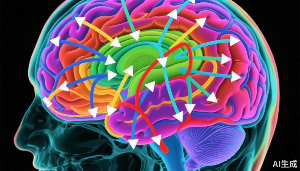
– Effective connectivity (EC) analysis between cerebellum and fronto-temporal brain regions provides robust biomarkers for major depressive disorder (MDD) classification.
– Multi-site resting-state fMRI data processed with Granger causality and ComBat harmonization enables cross-site generalizability.
– Machine learning classification using LightGBM achieved over 94% accuracy, sensitivity, and specificity in both cross-validation and independent validation.
– This approach offers a quantitative, objective adjunct to traditionally subjective MDD diagnostic methods.
Study Background
Major Depressive Disorder (MDD) remains a leading cause of disability worldwide, with profound impacts on quality of life and societal productivity. Diagnosis currently depends heavily on subjective clinical interviews and patient self-reports, which can be influenced by reporting biases and lack objective neurobiological correlates. There is a critical unmet need for reliable biomarkers that complement clinical assessments to improve diagnostic accuracy and facilitate earlier intervention.
Resting-state functional magnetic resonance imaging (rs-fMRI) has emerged as a powerful tool to investigate intrinsic brain network dynamics without task-based confounds. Specifically, effective connectivity (EC) analysis assesses the directionality and influence among brain regions, offering mechanistic insights beyond mere correlation. Previous studies have implicated dysfunction within frontal and temporal cortices, as well as the cerebellum, in MDD pathophysiology. However, translation of these findings into diagnostic tools has been limited by small sample sizes and lack of multi-site validation.
Study Design and Methods
This investigation employed a large multi-site rs-fMRI dataset of patients diagnosed with MDD and healthy controls. The authors applied Granger causality analysis to extract EC features reflective of directed neural interactions between brain regions, focusing on the cerebellum and fronto-temporal areas known to be implicated in affect regulation.
To address inter-site variability, the ComBat harmonization algorithm was used to adjust the EC features, ensuring that site-related differences did not confound the analysis. Additionally, multivariate linear regression controlled for demographic covariates such as age and sex.
Discriminative EC features were identified through statistical testing (two-sample t-tests) and refined via a model-based feature selection approach. The Light Gradient Boosting Machine (LightGBM), a powerful machine learning algorithm, was employed for classification of MDD versus healthy controls.
Rigorous model validation was performed using nested five-fold cross-validation and further tested in an independent external validation dataset to assess generalizability.
Key Findings
The analysis revealed 97 EC features primarily involving effective connectivity between the cerebellum and fronto-temporal regions as highly discriminative of MDD status. This aligns with accumulating evidence suggesting the cerebellum’s role in mood regulation and cognitive-affective integration via its connections with prefrontal and temporal cortices.
The classification model demonstrated exceptional performance with an overall accuracy of 94.35% during internal cross-validation, sensitivity of 93.52%, and specificity of 95.25%. When applied to the independent dataset, it retained accuracy of 94.74%, sensitivity of 90.59%, and specificity of 96.75%, confirming robust generalizability across different populations and imaging sites.
These results underscore the diagnostic potential of EC features derived from rs-fMRI as objective biomarkers for MDD, which significantly exceed typical accuracies reported in previous neuroimaging classification studies that often range between 70-85%.
Expert Commentary
The incorporation of effective connectivity metrics provides a deeper mechanistic understanding of circuit-level dysfunction in MDD compared to traditional functional connectivity measures. By capturing the direction of information flow, EC elucidates the dynamic pathological interactions driving depressive symptomatology.
The use of multisite data and harmonization techniques like ComBat addresses the critical issue of scanner and site variability, which historically hampers replication efforts in neuroimaging biomarker research. This methodological rigor enhances the translational value of the findings.
Nonetheless, despite high classification performance, it is important to consider that such models require validation in prospective clinical cohorts and across diverse demographic groups to fully ascertain their utility in routine practice. Moreover, longitudinal studies are necessary to determine whether these EC signatures can track treatment response or predict relapse.
Conclusion
This study compellingly demonstrates that effective connectivity between the cerebellum and fronto-temporal brain regions derived from resting-state fMRI serves as a powerful biomarker for the objective identification of major depressive disorder. The combination of advanced connectivity analysis, machine learning, and multi-site harmonization highlights a promising path toward integrating neuroimaging-based tools into psychiatric diagnostics.
Future research should focus on validating these findings in clinical settings, exploring the potential for monitoring disease progression and treatment efficacy, and simplifying acquisition pipelines for broader accessibility. Ultimately, the integration of neurobiological metrics such as EC could facilitate earlier, more precise diagnosis and personalized therapeutic strategies for MDD.
Funding and Clinical Trials
The original study did not specify funding sources or clinical trial registration information.
References
Dai P, Huang K, Shi Y, Xiong T, Zhou X, Liao S, Huang Z, Yi X, Grecucci A, Chen BT. Effective connectivity between the cerebellum and fronto-temporal regions correctly classify major depressive disorder: fMRI study using a multi-site dataset. J Affect Disord. 2025 Dec 1;390:119783. doi: 10.1016/j.jad.2025.119783. Epub 2025 Jul 1. PMID: 40609648.


