Introduction and Context
Neoadjuvant therapy — treatment given before surgery — is increasingly used to downstage tumors, improve resectability, and test systemic agents in the pre‑operative window. In several cancers (breast, rectal, bladder), the degree of pathological response after neoadjuvant therapy (for example, the proportion of residual viable tumor or a pathological complete response) is an accepted surrogate of treatment efficacy and can predict outcome. Until now, renal cell carcinoma (RCC) has lacked an international, standard approach for how to prepare nephrectomy specimens and how to measure and report pathological response after pre‑operative systemic therapy.
In a systematic review and consensus effort published by the International Neoadjuvant Kidney Cancer Consortium (INKCC) led by Blackmur et al. (Lancet Oncology, 2025), investigators reviewed the reporting practices for neoadjuvant RCC and produced practical, consensus recommendations to standardize tissue handling and reporting in trials and research settings. The guideline responds to several clinical gaps: heterogeneous reporting in the literature, inconsistent sampling that limits reproducibility, and lack of agreed definitions for what constitutes a meaningful pathological response in RCC.
Why this matters now
– Systemic therapy options for RCC have evolved rapidly: immune checkpoint inhibitors (ICIs), combination ICI/tyrosine kinase inhibitor (TKI) regimens, and newer targeted agents are increasingly tested in earlier disease and peri‑operative settings. Reliable pathological endpoints are needed to compare agents and potentially accelerate drug development.
– Existing pathology protocols for nephrectomy (for diagnosis and staging) do not provide a unified method for assessing treatment effect after neoadjuvant therapy.
– The INKCC review found major heterogeneity: among 119 studies of pre‑surgical therapy in RCC, only five reported how pathological response was assessed; qualitative statements predominated, and only eight studies used quantitative assessment (Blackmur et al., Lancet Oncol. 2025).
New Guideline Highlights
Main goals of the INKCC recommendations
– Provide a standardized baseline sampling approach for nephrectomy specimens after neoadjuvant therapy.
– Define how to quantify and report residual viable tumor in a reproducible manner.
– Recommend clinical data elements to accompany pathology reports so pathological response can be interpreted in context.
– Encourage adoption in clinical trials and research while recognizing that links between pathological response metrics and long‑term outcomes require validation.
Key takeaways for clinicians and investigators
– Standardize gross sampling using a baseline template and consider additional sampling for large or heterogeneous tumors.
– Report residual viable tumor as both a percentage (in 10% intervals) and the greatest linear dimension of residual viable tumor.
– Document treatment regimen, number of cycles/doses, interval from last dose to surgery and imaging response.
– Use standardized terminology and clearly state if the assessment is trial‑driven research vs routine diagnostic reporting.
Updated Recommendations and Key Changes (Compared with Usual Practice)
Summary of what the INKCC consensus adds to current practice
– From qualitative to quantitative: Move away from vague terms such as “marked treatment effect” or “extensive necrosis” without quantification. The guideline recommends numerical reporting.
– Sampling template: Introduces a defined baseline sampling approach for nephrectomy specimens excised after neoadjuvant therapy.
– Dual metrics: Recommends reporting both percent residual viable tumor (in 10% increments) and greatest linear extent (in millimetres) of viable tumor, acknowledging both metrics capture different aspects of response.
– Clinical linkage: Requires specific clinical metadata in pathology reports to ensure reproducible interpretation across studies.
Why these changes? Evidence and rationale
– The systematic review showed rare quantitative reporting; inconsistency precludes pooled analyses and comparison across trials.
– Percent viable tumor mirrors approaches in other tumor types (e.g., lung, soft tissue) and provides an intuitive measure of tumor kill. Greatest linear extent acts as a size metric that can be correlated with radiologic measurements and staging.
– Standardized sampling reduces bias from focal residual tumor and heterogeneity, improving the robustness of pathological endpoints.
Topic-by-Topic Recommendations
Note: The INKCC consensus is intended primarily for clinical trials and research settings where pathological response is a predefined endpoint. Many recommendations are consensus‑based; formal graded evidence levels are not assigned because prospective validation is limited. Wherever possible, the guideline references existing pathology and oncology standards.
1) Pre‑analytic and clinical data to accompany the specimen
Essential clinical data to provide with the specimen:
– Neoadjuvant regimen (drug names, doses, start/stop dates).
– Number of cycles/treatments and dates of last dose before surgery.
– Pre‑treatment and pre‑operative radiologic tumor measurements (largest diameter, tumor volume if available).
– Clinical indication for nephrectomy (partial vs radical, urgency).
– Relevant prior treatments (e.g., embolization, local ablation).
Why: Histologic changes after therapy are interpreted in light of timing and type of therapy — especially where ICIs can produce atypical histologic features (inflammation, fibrosis) that mimic treatment effect.
2) Grossing and baseline sampling approach
Baseline template (minimum standard):
– For tumors ≤7 cm: sample the tumor at 1 block per centimeter of greatest tumor diameter (minimum 3 blocks).
– For tumors >7 cm: at least 1 block per centimeter up to 10 cm, then 1 additional block per 2 cm beyond 10 cm (or a predefined maximum to ensure feasibility).
– Mandatory blocks: tumor‑rim interface; areas of apparent necrosis; any suspicious satellite nodules; tumor‑venous thrombus if present.
– Photograph/capture gross images and map sampled areas.
Rationale: Neoadjuvant therapy often produces heterogeneous effects; more intensive, systematic sampling reduces the risk of missing residual viable tumor.
3) Microscopic assessment and reporting elements
Essential microscopic data elements (to be reported in the pathology report):
– Percent residual viable tumor (estimate in 10% increments: 0%, 1–10%, 11–20% … 91–100%). Predefined bins allow consistent categorization across reviewers and studies.
– Greatest linear dimension (GLD) in millimeters of the largest contiguous focus of viable tumor.
– Presence and distribution of therapy‑related changes (fibrosis, necrosis, hyalinization, inflammation) — qualitative descriptors permitted.
– Lymphovascular invasion and margin status (if applicable) — report using standard CAP criteria (College of American Pathologists).
– Tumor type and grade (WHO/ISUP nucleolar grade) assessed on viable tumor.
Pathological complete response (pCR): defined as no residual viable tumor identified in the sampled tumor bed and tumor‑bearing tissue. pCR must be explicitly stated and requires adequate sampling per the baseline template.
Why percent + GLD? Percent gives an index of therapy effect; GLD links to radiology and staging and may be more reproducible when small foci remain.
4) Special situations and practical considerations
– Partial nephrectomy specimens: apply the same principles, emphasizing detailed sampling of the tumor bed and margins.
– Multifocal tumors: assess each tumor separately and report aggregate and dominant tumor metrics.
– Tumor thrombus: sample thrombus thoroughly and report residual viable tumor within thrombus as a separate metric.
– Prior local therapies (embolization, cryoablation): note that morphologic changes may confound interpretation and require careful clinicopathologic correlation.
5) Data capture and trial use
– Pathology CRFs (case report forms) should include the recommended elements to facilitate pooled analyses.
– Digital pathology and central review are encouraged for multi‑center trials to harmonize assessments.
Recommendation Grades and Evidence
– The INKCC guidance is a consensus statement based on systematic review and expert workshop deliberations rather than a formal, graded guideline backed by large randomized evidence.
– Recommendations are pragmatic, evidence‑informed and designed to harmonize reporting; prospective studies are needed to correlate these pathological metrics with survival outcomes (disease‑free and overall survival).
Example quick reference (recommended minimal dataset for trials):
– Clinical: regimen, last dose date, imaging metrics.
– Grossing: photograph, map, sampling per baseline template.
– Microscopy: percent residual viable tumor (10% bins), greatest linear dimension (mm), pCR yes/no, margins, lymphovascular invasion, grade of residual tumor.
Expert Commentary and Insights
Consensus panel perspectives
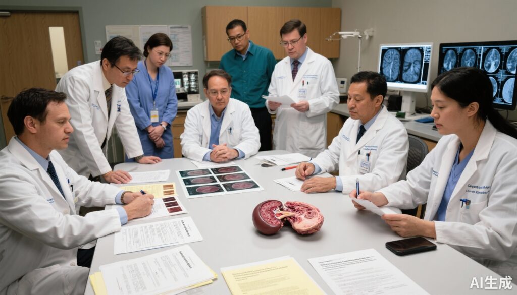
– Harmonization is a priority: Several experts emphasized that without standardization, trials will continue to report incomparable pathological endpoints. The workshop at the Netherlands Cancer Institute (INKCC) concluded that a modest increase in sampling burden is justified by the gains in reliability.
– Pragmatism vs perfection: The group acknowledged trade‑offs between exhaustive sampling and practical feasibility in routine diagnostic practice; the recommendations are therefore pitched primarily at the trial/research setting, with potential adoption into routine practice after validation.
– Quantitative bins: Using 10% intervals strikes a balance between precision and interobserver reproducibility.
Controversies and open questions
– Which metric best predicts outcome? Percent viable tumor vs absolute GLD — this remains unresolved and requires pooled analyses with survival endpoints.
– Role of immune‑related histologic patterns: ICIs can produce pronounced lymphoid infiltrates and fibrosis that may not equate to tumor cell death; standardized descriptors and possibly ancillary studies (e.g., immunohistochemistry) might be needed in some settings.
– Central review vs local reporting: Central review increases consistency but adds logistical complexity; the panel recommends central review for pivotal trials.
Future research priorities identified by the panel
– Prospective validation of pathological response metrics against relapse‑free and overall survival in neoadjuvant trials.
– Interobserver reproducibility studies to test percentage bins and GLD measurements.
– Investigations into biomarkers (tissue and blood) that correlate with pathological response to identify responders earlier.
Practical Implications for Daily Practice
For trialists and pathologists
– Trial designers should incorporate the INKCC recommended dataset in protocols and CRFs.
– Pathology labs participating in neoadjuvant RCC trials should train staff on the baseline sampling template, include appropriate gross photography and ensure the required clinical metadata travel with the specimen.
– Consider central pathology review for registration‑intended trials or when pathology is a key endpoint.
For surgeons and oncologists
– Provide detailed treatment timelines and pre‑operative imaging with specimens to support accurate pathologic interpretation.
– Understand that pathological response assessments are evolving endpoints; their predictive value for patient outcomes in RCC will be defined over the coming years as prospective data accumulate.
Patient vignette: Applying the guideline
Michael, a 63‑year‑old man, received 3 cycles of neoadjuvant ICI/TKI combination for a 9‑cm right renal mass before planned radical nephrectomy. At surgery, the specimen was photographed and sampled using the INKCC baseline template (1 block/cm). The pathology report recorded: percent residual viable tumor 20% (10–30% bin), greatest linear dimension of residual viable tumor 18 mm, histologic subtype clear cell RCC, WHO/ISUP grade 3 on residual tumor, negative margins, and no viable tumor in sampled thrombus. These standardized data allowed the trial team to classify his pathological response and included him in a pooled analysis correlating pathological metrics with 2‑year relapse‑free survival.
References
1. Blackmur JP, van der Mijn JCK, Warren AY, Browning L, Burgers F, Hirsch MS, Kapur P, Mehra R, Rao P, Signoretti S, Bex A, Stewart GD, van Montfoort ML, Jones JO; International Neoadjuvant Kidney Cancer Consortium. Assessing pathological response to neoadjuvant therapy in renal cell carcinoma: a systematic review and guidelines for sampling and reporting standards from the International Neoadjuvant Kidney Cancer Consortium. Lancet Oncol. 2025 Oct;26(10):e536-e546. doi:10.1016/S1470-2045(25)00345-6.
2. National Comprehensive Cancer Network. NCCN Clinical Practice Guidelines in Oncology: Kidney Cancer. (Accessed 2024–2025 versions.) Available at: https://www.nccn.org
3. European Association of Urology Guidelines on Renal Cell Carcinoma. EAU Guidelines. (2024 edition). Available at: https://uroweb.org/guideline/renal-cell-carcinoma/
4. College of American Pathologists. Protocol for the Examination of Nephrectomy Specimens for Renal Cell Carcinoma. CAP Cancer Protocols. (Latest version available online.) https://www.cap.org
5. Amin MB, Edge SB, Greene FL, et al. AJCC Cancer Staging Manual, 8th edition. Springer; 2017.
6. WHO Classification of Tumours Editorial Board. WHO Classification of Tumours: Urinary and Male Genital Tumours, 5th Edition. IARC; 2022.
7. International Collaboration on Cancer Reporting (ICCR). Dataset for Reporting Renal Cell Carcinoma; recommended core items. (Accessed 2024.) https://www.iccr-cancer.org
Conclusion
The INKCC consensus provides a practical, standardized framework to quantify pathological response in RCC after neoadjuvant therapy. The recommendations — standardized sampling, quantification of residual viable tumor in 10% increments plus greatest linear extent, and mandatory clinical metadata — are designed to make pathological endpoints reliable and comparable across trials. Their adoption in clinical trials will help determine whether pathological response can become a validated surrogate of long‑term outcomes in renal cancer and may, in time, influence routine reporting practice. For now, these guidelines are a key step towards rigorous, reproducible evaluation of neoadjuvant strategies in RCC.


