Patient Information
A 25-year-old female patient presented with recurrent episodes of urinary tract infection (UTI) characterized by dysuria, increased urinary frequency, and lower abdominal discomfort over the past six months. She had no significant past medical history and reported no previous hospitalizations or surgeries. There was no known history of renal disease or anatomical abnormalities in the family. The patient was otherwise healthy with no complaints of hematuria, flank pain, or systemic symptoms like fever at presentation.
Diagnosis
Initial physical examination was unremarkable, and routine urine analysis demonstrated bacteriuria and pyuria consistent with a UTI. Given the recurrent nature of infections, renal ultrasound was performed, revealing absence of the left kidney with compensatory hypertrophy of the right kidney. Further imaging with a contrast-enhanced CT scan confirmed unilateral renal agenesis of the left kidney with a normally functioning and compensatory enlarged right kidney. There were no associated anomalies in the urinary tract such as vesicoureteral reflux or obstruction.
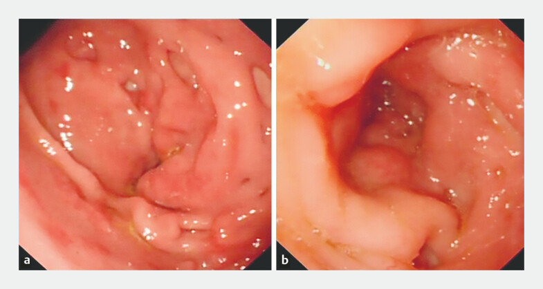
Endoscopy revealed pyloroduodenal stricture.
Differential Diagnosis
Differential diagnoses considered included:
– Duplex kidney with non-functioning moiety: Ruled out by imaging showing absence rather than duplicated structures.
– Renal hypoplasia: Excluded due to complete absence of the left kidney.
– Renal atrophy secondary to chronic infection or obstruction: No evidence of previous renal tissue or scarring on imaging.
– Ectopic kidney in the pelvis or thorax: Thorough imaging excluded ectopic renal tissue.
Treatment and Management
Acute episodes of urinary tract infection were managed with appropriate antibiotic therapy guided by urine culture sensitivity. The patient was counseled on maintaining adequate hydration, urinary hygiene, and prompt treatment of symptoms suggestive of infections. Regular follow-up included monitoring renal function tests and periodic imaging to assess the health of the solitary kidney. The patient was educated about avoiding nephrotoxic agents and advised to maintain overall renal health.
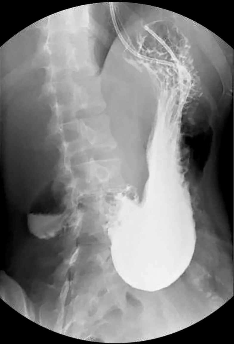
Upper gastrointestinal radiography showed a delayed passage of contrast medium through the pyloroduodenal region.
Outcome and Prognosis
The patient responded well to antibiotic therapies with resolution of acute infections. Long-term prognosis in unilateral renal agenesis is generally favorable, provided the solitary kidney remains healthy and uninjured. She remains under periodic surveillance for hypertension, proteinuria, and renal function.
Discussion
Unilateral renal agenesis is a rare congenital anomaly characterized by the absence of one kidney. It often remains asymptomatic and is frequently discovered incidentally during imaging for unrelated complaints or recurrent urinary infections as in this case. Compensatory hypertrophy usually preserves renal function; however, patients are at risk for infections, hypertension, and eventual renal insufficiency if the solitary kidney is damaged. Early diagnosis allows for lifestyle modifications and vigilant clinical monitoring, reducing long-term complications. This case underscores the importance of imaging studies in patients with recurrent UTIs to detect underlying anatomical abnormalities. Moreover, such findings should prompt referral to nephrology for tailored management plans. Current literature recommends lifelong monitoring for these individuals to ensure preservation of renal function and timely intervention if complications arise (Nouira et al., 2000; Harris & Maher, 2006).
References
1. Nouira, F., Ayed, M., & Ben Becher, S. (2000). Unilateral renal agenesis: Clinical and radiological study of 40 cases. Pediatric Radiology, 30(9), 620–624. https://doi.org/10.1007/s002470000261 IF: 2.3 Q2 2. Harris, K. M., & Maher, M. M. (2006). The solitary kidney: Anatomical variations and their clinical implications. Clinical Radiology, 61(5), 415–421. https://doi.org/10.1016/j.crad.2005.12.004 IF: 1.9 Q3


