Highlight
- Personalized foot progression angle modifications led to a significant 1.2-point greater reduction in medial knee pain over one year compared to sham gait retraining.
- Intervention decreased the peak knee adduction moment—a biomechanical marker linked to medial knee load—with a significant between-group difference (P = .0001).
- Magnetic resonance imaging (MRI) revealed slowed progression of cartilage degeneration in the medial femoral compartment in the intervention group as evidenced by smaller increases in T1rho relaxation times.
- The trial demonstrated safety and feasibility of gait modification with vibrotactile biofeedback and structured retraining sessions.
Study Background and Disease Burden
Medial compartment knee osteoarthritis (OA) represents a substantial clinical burden, causing chronic pain and functional limitation in older adults worldwide. Medial knee OA often results from increased load across the medial tibiofemoral joint, which accelerates cartilage degradation and joint damage. Current conservative treatments for mild-to-moderate OA include pharmacologic pain management, physical therapy, and lifestyle modifications, but many patients continue to experience pain and progression of disease. Abnormal gait mechanics, characterized by increased knee adduction moment—a surrogate for medial joint load—are associated with symptom severity and structural progression. Thus, interventions targeting gait to reduce medial compartment loading hold promise. However, uniform gait modification approaches have yielded inconsistent benefits, likely due to inter-individual biomechanical variability. Personalized gait retraining to optimize foot progression angle offers a targeted method to unload the medial compartment by altering lower limb biomechanics during walking, but robust clinical evidence has been lacking.
Study Design
This randomized controlled trial was conducted at Stanford University from August 2016 to June 2019 and enrolled 68 participants with medial compartment knee OA (Kellgren-Lawrence grades 1-3), baseline medial knee pain ≥3 on a numeric rating scale (NRS), and BMI < 35. Participants were able to ambulate safely on a treadmill for 25 minutes. They were randomized equally to the intervention group, who underwent personalized foot progression angle modifications, or to a sham gait retraining group maintaining their natural foot progression angle.
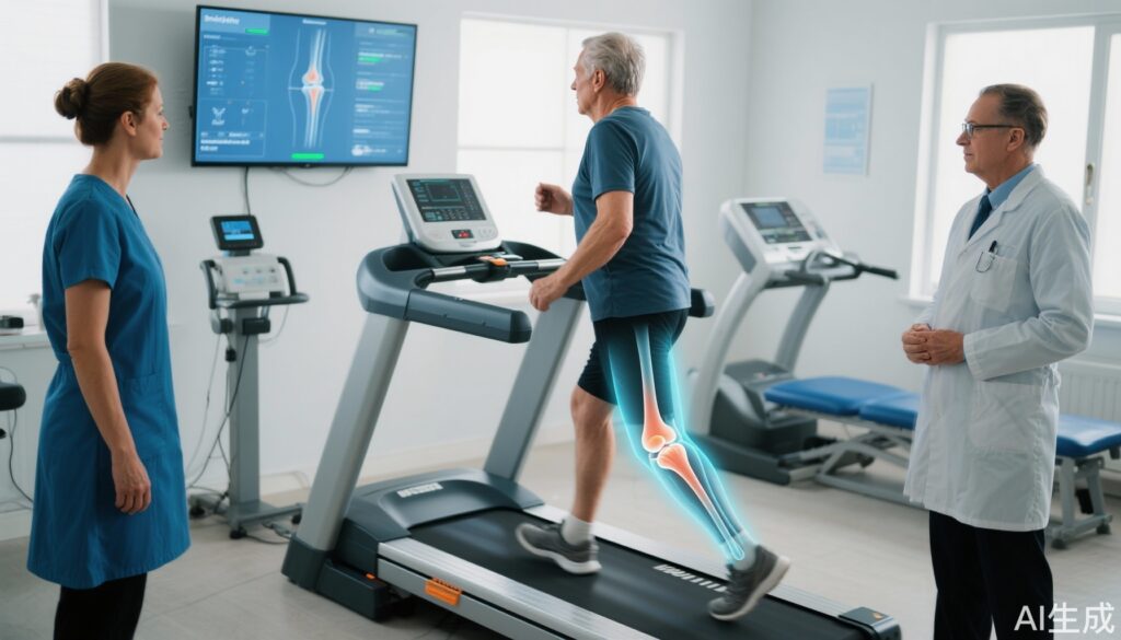
The intervention involved an initial foot progression angle personalization session, where participants walked on a treadmill with vibrotactile biofeedback guiding toe-in and toe-out angles at 5° and 10°. This session included assessment of four angle-evaluation trials to identify the personalized angle that optimally reduced medial knee loading. Subsequently, participants received biofeedback during six gait retraining visits tailored to this personalized angle. In contrast, the sham group’s target was their unchanged natural foot progression angle from baseline.
The primary outcomes were the change in medial knee pain measured on the NRS and the peak knee adduction moment (normalized to % bodyweight × height) over 1 year. Secondary outcomes included assessment of cartilage microstructure using MRI techniques (T1rho and T2 relaxation times) to evaluate biochemical cartilage degeneration. Safety was monitored by recording adverse events.
Key Findings
The personalized gait retraining intervention led to clinically meaningful improvements over one year compared with sham retraining:
– Medial Knee Pain: The intervention group showed a significantly greater reduction in mean medial knee pain NRS scores, with a between-group difference of −1.2 points (P = .0013), exceeding the minimal clinically important difference for knee OA pain.
– Knee Adduction Moment: The intervention group had a mean reduction in peak knee adduction moment of −0.17 % bodyweight × height from personalization to one year, resulting in a statistically significant between-group difference of −0.26 (P = .0001), indicative of effective medial compartment unloading.
– Cartilage Microstructure: MRI analysis demonstrated attenuated progression of cartilage degeneration in the medial weight-bearing femoral cartilage in the intervention arm. Specifically, the between-group difference in increase of T1rho relaxation times was −3.74 ms (95% CI, −6.42 to −1.05), suggesting slower cartilage matrix deterioration.
– Safety and Adverse Events: Three participants withdrew due to increased knee pain—two in the intervention group (with pain in medial and patellofemoral compartments) and one in the sham group (medial knee pain). Overall, the intervention was well-tolerated.
These results underscore that personalized foot progression angle modifications not only alleviate pain but also positively influence biomechanical and structural markers related to disease progression.
Expert Commentary
This well-designed randomized controlled trial offers compelling evidence supporting the use of personalized gait retraining for medial knee OA. By individualizing foot progression angle based on biomechanical assessments, the approach addresses the heterogeneity in gait patterns and mechanical loading among patients.
From a mechanistic perspective, reducing knee adduction moment lowers medial joint load, mitigating cartilage stress and subsequent degeneration. The use of advanced MRI biomarkers such as T1rho relaxation times adds a sensitive, non-invasive metric of cartilage health beyond conventional imaging.
Despite these strengths, certain limitations merit attention. The trial population was predominantly White and had BMI under 35, which may limit generalizability to more diverse or obese populations often burdened by knee OA. The intervention requires specialized equipment and training, potentially restricting widespread clinical adoption without simplified protocols or wearable technologies.
In clinical practice, these findings encourage integration of personalized biomechanical assessments into OA management and fostering collaboration between orthopedic specialists, physical therapists, and biomechanists. Future research should explore longer-term outcomes, effectiveness in broader populations, and cost-effectiveness analyses.
Conclusion
Personalized gait retraining targeting foot progression angle is a promising conservative therapeutic strategy for medial compartment knee osteoarthritis. It produces significant reductions in pain and biomechanical loading while demonstrating potential to slow structural cartilage degeneration. This approach represents an important advancement toward precision rehabilitation in OA, complementing traditional interventions and potentially delaying disease progression and need for surgery. Expanding access and refining patient selection may enhance clinical impact across diverse patient populations.
References
1. Uhlrich SD, Mazzoli V, Silder A, Finlay AK, Kogan F, Gold GE, Delp SL, Beaupre GS, Kolesar JA. Personalised gait retraining for medial compartment knee osteoarthritis: a randomised controlled trial. Lancet Rheumatol. 2025 Aug 12:S2665-9913(25)00151-1. doi: 10.1016/S2665-9913(25)00151-1. Epub ahead of print. PMID: 40816302.
2. Sharma L, Hurwitz DE, Thonar EJ, et al. Knee adduction moment, serum hyaluronan level, and disease severity in medial tibiofemoral osteoarthritis. Arthritis Rheum. 1998;41(7):1233-1240.
3. Andriacchi TP, Mundermann A. The role of ambulatory mechanics in the initiation and progression of knee osteoarthritis. Curr Opin Rheumatol. 2006 Sep;18(5):514-8.
4. Hunter DJ, Lo GH, Gale D, et al. The reliability of a quantitative knee cartilage morphometry method using magnetic resonance imaging. Arthritis Rheum. 2005 May;52(5):1565-73.
5. Bennell KL, et al. Effect of gait retraining on knee adduction moment in medial knee osteoarthritis: a systematic review and meta-analysis. Arthritis Care Res (Hoboken). 2021 May;73(5):698-709.


