Study Background and Disease Burden
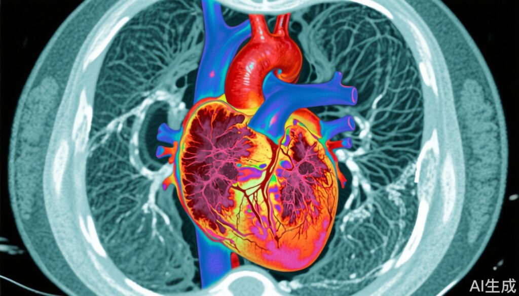
Heart failure (HF) represents a significant global health challenge, affecting millions worldwide with high morbidity and mortality. It encompasses a spectrum of conditions characterized by the inability of the heart to pump efficiently, leading to insufficient perfusion of organs. Underlying myocardial fibrosis plays a critical role in the progression and adverse outcomes of HF by contributing to myocardial stiffness, impaired contractility, and arrhythmogenic substrate formation. Traditionally, myocardial fibrosis was assessed invasively by biopsy, but this poses risks and is limited by sampling error. Noninvasive cardiac magnetic resonance (CMR) techniques, notably T1 mapping and extracellular volume fraction (ECV) measurement, have emerged as pivotal tools for quantitative characterization of diffuse myocardial fibrosis.
Non-ischaemic dilated cardiomyopathy (NIDCM), a leading cause of HF with reduced ejection fraction (HFrEF), also carries significant risks of heart failure progression and arrhythmic events. Understanding how T1 mapping and ECV relate to adverse events in NIDCM patients is essential for risk stratification and personalized therapeutic approaches.
Study Design
Two recent meta-analyses comprehensively evaluated the prognostic value of myocardial T1 mapping and ECV in distinct HF populations. The first study by Zhang et al. examined the entire HF spectrum, including both HF with reduced ejection fraction (HFrEF) and HF with preserved ejection fraction (HFpEF), incorporating 19 studies totaling 5,384 patients. Searches were conducted in PubMed, Web of Science, and SCOPUS to identify studies reporting baseline native myocardial T1 and ECV values and their association with clinical outcomes such as mortality and HF-related events. Hazard ratios (HRs) and mean differences were pooled, with subgroup analyses by HF type and clinical factors.
The second meta-analysis by Marchini et al. focused on 12 observational studies involving 4,025 patients with non-ischaemic dilated cardiomyopathy. The primary endpoint was major adverse cardiovascular events (MACE), including heart failure-related and arrhythmic events. The study assessed the prognostic significance of native T1 and ECV using random-effects meta-analysis, reporting pooled HRs and weighted mean differences between those experiencing events and those without.
Key Findings
In Zhang et al.’s meta-analysis, patients experiencing adverse outcomes had significantly elevated native myocardial T1 and ECV values compared to those without events, with weighted mean differences of 41.17 ms for native T1 and 4.73% for ECV. Both markers showed consistent positive associations with endpoints throughout the HF population. Specifically, a 1% increase in ECV conferred a 20% higher risk of adverse events (HR 1.20, 95% CI 1.13–1.28), and larger increments were associated with proportionally increased risk (HR binary 2.62). Native T1 similarly demonstrated risk elevation (HR per 1-ms increase: 1.02; HR binary: 2.93). However, in HFpEF, native T1’s predictive value was not statistically significant, whereas ECV retained prognostic utility across HF subtypes. Subgroups of younger patients with severe cardiac insufficiency (NYHA III-IV), those with increasing ECV over time, or those without detectable late gadolinium enhancement but abnormal T1 mapping were at particularly high risk.
Marchini et al.’s results in NIDCM showed that both native T1 and ECV significantly predicted MACE. The pooled HR was 1.07 per unit increase in native T1 (95% CI 1.04–1.09) and 1.37 per unit increase in ECV (95% CI 1.29–1.44). For secondary endpoints, including heart failure and arrhythmia, T1 and ECV remained significant predictors. Patients with events showed higher native T1 (mean difference 30.91 ms) and ECV (mean difference 4.52%) than those without. Follow-up durations averaged 22 months, supporting the robust short-to-mid-term prognostic utility.
Expert Commentary
These meta-analyses reinforce myocardial fibrosis as a key driver of adverse outcomes in HF and NIDCM and validate CMR T1 mapping and ECV as integral quantitative biomarkers. The demonstrated superiority of ECV over native T1 in prognostic performance, especially in HFpEF where native T1 was less predictive, may relate to ECV specifically reflecting extracellular matrix expansion and fibrosis, while native T1 can be influenced by multiple factors including edema or infiltration.
Clinically, ECV measurement provides a reliable, reproducible quantitative assessment that can be standardized across centers, promoting its incorporation into routine risk stratification algorithms. The subgroup findings highlight the need for targeted surveillance in younger patients with severe symptoms and those with persistent ECV elevation, who may benefit from intensified therapies or closer monitoring.
Nonetheless, limitations include heterogeneity in CMR protocols and population characteristics across studies. While meta-analytic approaches statistically reconcile variability, future large-scale prospective cohorts with uniform imaging protocols and longer follow-up would enhance generalizability. Importantly, these imaging biomarkers should be integrated with clinical indices and other modalities for comprehensive management.
Conclusion
Myocardial T1 mapping and ECV fraction via cardiac MRI are emerging as powerful, noninvasive prognostic tools in heart failure across the spectrum of ejection fraction and in non-ischaemic dilated cardiomyopathy. ECV consistently predicts adverse cardiovascular events, outperforming native T1 particularly in HFpEF. Incorporation of these advanced imaging biomarkers into clinical practice holds promise for improving risk stratification, guiding personalized therapy, and ultimately improving outcomes in patients with HF and NIDCM. Ongoing research and consensus efforts will be vital to optimize their clinical implementation and elucidate mechanistic links to myocardial fibrosis and remodeling.
References
1. Zhang H, Yang W, Zhou D, et al. Prognostic Value of Myocardial T1 Mapping and Extracellular Volume Fraction in Heart Failure: A Meta-Analysis. JACC Cardiovasc Imaging. 2025 Aug 28:S1936-878X(25)00369-9. doi: 10.1016/j.jcmg.2025.06.017. Epub ahead of print. PMID: 40884516.
2. Marchini F, Dal Passo B, Campo G, et al. T1 mapping and major cardiovascular events in non-ischaemic dilated cardiomyopathy: a systematic review and meta-analysis. ESC Heart Fail. 2025 Aug;12(4):2621-2630. doi: 10.1002/ehf2.15279. Epub 2025 Apr 25. PMID: 40285366; PMCID: PMC12287780.