Highlight
– Excess supplemental iron intake is linked to accelerated phenotypic aging via inflammatory pathways.
– A U-shaped relationship exists between total iron intake and aging, with a protective effect below a threshold and harmful effects beyond it.
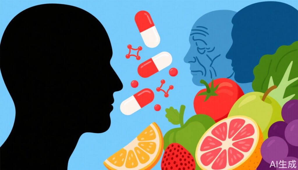
– Dietary iron alone does not accelerate aging, highlighting risks associated with supplement-driven iron overload.
– Anti-inflammatory and antioxidant-rich diets can counteract the pro-aging effects of iron overload.
– Inflammation mediates a significant portion (15.5%-25.6%) of the aging acceleration associated with excess iron supplementation.
Study Background and Disease Burden
Iron is an essential trace element critical for numerous biological processes including oxygen transport, DNA synthesis, and cellular respiration. However, both iron deficiency and overload pose significant health risks. With aging populations worldwide, understanding modifiable factors influencing biological aging is imperative. Phenotypic age, a composite biomarker reflecting biological rather than chronological aging, predicts morbidity and mortality risks more accurately than chronological age alone. Recent concerns have arisen around excessive iron intake from dietary supplements common in many populations, implicating potential adverse effects on aging through mechanisms including inflammation and oxidative stress. Intriguingly, iron has dual effects: it is indispensable but can catalyze reactive oxygen species (ROS) formation, promoting oxidative damage when present in excess. Despite recognition of these dual roles, the relationship between iron intake from various sources and phenotypic aging, and the roles of inflammatory or oxidative pathways in this relationship, remain poorly elucidated.
Study Design
This cross-sectional analysis utilized data from the National Health and Nutrition Examination Survey (NHANES) 2017-2018 cycle, comprising 8692 participants aged 20 years or older. Iron intake was quantified from both dietary sources and supplements. The primary endpoint was phenotypic age acceleration (PhenoAgeAccel), derived from validated biomarker algorithms. Nonlinear associations between iron intake and PhenoAgeAccel were modeled using weighted restricted cubic splines (RCS). Multivariable-adjusted weighted generalized linear models (GLMs) adjusted for confounders were applied to assess effect sizes. K-means clustering identified co-exposure patterns to iron, segregating populations into groups based on iron intake profiles. Interaction effects with dietary anti-inflammatory or antioxidant potential were evaluated using likelihood ratio tests. To investigate mechanistic pathways, mediation analysis quantified the contribution of inflammation and oxidative stress biomarkers to the relationship between iron overload and phenotypic aging. Additional adjustments accounted for anemia status and post-menopausal status among women to control for potential confounders.
Key Findings
A robust U-shaped relationship between total iron intake and PhenoAgeAccel was observed, with a critical breakpoint at 18.441 mg/day. Below this threshold, increased iron intake was associated with protective effects against accelerated aging (β = -0.126, indicating a deceleration of aging), whereas intake above this breakpoint correlated with accelerated phenotypic aging (β = 0.021). Importantly, iron obtained solely from diet was not associated with aging acceleration, suggesting safety within typical dietary ranges.
Conversely, supplemental iron intake showed a significant positive association with PhenoAgeAccel (β = 0.017), indicating that excess iron from supplements may promote aging acceleration. The analysis revealed no significant gender interaction effects.
K-means clustering stratified participants into two distinct groups: the dietary iron reference (DIR) group, characterized by total iron intake predominantly below the established upper limit (UL) of 45 mg/day with minimal supplemental iron intake, and the supplement-driven iron overload (SDIO) group, all exceeding the UL threshold, with supplemental iron composing 83.44% of total iron intake on average. The SDIO group exhibited significantly faster phenotypic aging relative to the DIR group (β = 1.774, P < 0.05), underscoring the deleterious effects of supplement-driven iron overload.
Notably, participants consuming diets rich in anti-inflammatory or antioxidant properties showed a significant interaction effect, whereby such dietary patterns counteracted the iron-related acceleration of aging (P for interaction = 0.025). Mediation analyses demonstrated that inflammatory biomarkers partially mediated the detrimental impact of supplement-driven iron overload on accelerated aging, accounting for 15.53% to 25.63% of this effect. Oxidative stress markers showed a less pronounced mediation effect.
The robustness of these findings persisted after controlling for anemia and post-menopausal status, mitigating the risk of confounding due to these conditions.
Expert Commentary
This well-designed epidemiological study adds valuable insight into the complex role of iron intake on biological aging. Its use of a large, nationally representative cohort and sophisticated modeling lends strength to the findings. The identification of a U-shaped curve is biologically plausible, reflecting the narrow therapeutic window of iron—deficiency impairs physiological function, whereas excess incites oxidative and inflammatory damage.
The distinction between dietary iron and supplemental iron is clinically important. Supplemental iron bypasses physiological regulatory mechanisms, potentially leading to systemic iron overload. This is consistent with prior literature linking iron overload states to chronic inflammatory conditions and age-related pathologies such as cardiovascular disease and neurodegeneration.
The partial mediation by inflammation biomarkers underscores chronic inflammation, or “inflammaging,” as a central mechanism by which iron excess accelerates aging. This aligns with the broader scientific consensus recognizing systemic inflammation as a hallmark of aging.
Encouragingly, the study highlights the potential of anti-inflammatory and antioxidant diets to mitigate iron-induced aging acceleration, supporting nutritional interventions as feasible, low-risk strategies to preserve healthy aging. However, the cross-sectional design limits causal inference, and residual confounding cannot be excluded. Further longitudinal and interventional studies are warranted to delineate causality and test dietary modification strategies.
Conclusion
This study compellingly demonstrates that excessive iron intake from supplements, but not dietary sources, is associated with accelerated phenotypic aging, mediated in part by inflammatory pathways. Supplement-driven iron overload beyond established upper limits significantly increases biological aging velocity. However, adherence to anti-inflammatory or antioxidant-rich dietary patterns may counterbalance these adverse effects. Clinicians should caution against indiscriminate iron supplementation, particularly in populations without clinical deficiency, and emphasize balanced nutrition to promote healthy aging. Future research should explore targeted interventions to modulate iron homeostasis and inflammation in aging populations.
References
Li B, Ming Z, Wang Y, Zhang Y, Zhou R, Zhang C, Wu Y, Wang G, Xie H, Li Y, Li R. Supplement-driven iron overload accelerates phenotypic aging via inflammatory biomarkers: Potential counteraction through anti-inflammatory or antioxidant diets. Redox Biol. 2025 Sep;85:103733. doi: 10.1016/j.redox.2025.103733. Epub 2025 Jun 19. PMID: 40570516; PMCID: PMC12246624.
Ganz T. Iron homeostasis in host defence and inflammation. Nat Rev Immunol. 2016 Aug;16(8):566-78. doi: 10.1038/nri.2016.84.
Lopez-Otin C, Blasco MA, Partridge L, Serrano M, Kroemer G. The Hallmarks of Aging. Cell. 2013 Jun 6;153(6):1194-217. doi: 10.1016/j.cell.2013.05.039.
Calder PC, Ahluwalia N, Albers R, Bosco N, Bourdet-Sicard R, Haller D et al. A consideration of biomarkers to be used for evaluation of inflammation in human nutritional studies. Br J Nutr. 2013 Aug 28;109 Suppl 1:S1-34. doi: 10.1017/S0007114512005115.