Metformin kích hoạt Ferroptosis trong AML thông qua tái cấu trúc lipid: Cơ hội tái sử dụng cho các tiểu loại chuyển hóa

Metformin kích hoạt Ferroptosis trong AML thông qua tái cấu trúc lipid: Cơ hội tái sử dụng cho các tiểu loại chuyển hóa

Nhấn mạnh

– Metformin gây ra các loài phản ứng oxy (ROS) và cái chết tế bào ferroptosis trong các mẫu AML cấp tính in vitro, với hiệu quả lớn nhất ở các trường hợp có chuyển hóa lipid bị gián đoạn (đặc biệt là bệnh đột biến IDH2 và FLT3).

– Việc tái cấu trúc lipid với tăng triglycerides và axit béo đa không no (PUFAs), điều hòa tăng các gen hạt lipid bao gồm DGAT1, và sự hấp thụ thông qua CD36 là những yếu tố quyết định chính của độ nhạy cảm đối với metformin.

– Sự ức chế DGAT1 tạo ra sự hiệp đồng với metformin; các chất chelate sắt làm giảm tác dụng của nó—ủng hộ ferroptosis là cơ chế cái chết tế bào hoạt động và chỉ ra các chiến lược kết hợp hợp lý.

Nền tảng

Sự lập trình chuyển hóa là một đặc điểm nổi bật của ung thư và là một điểm yếu điều trị được nhận biết trong bệnh bạch cầu cấp tính myeloid (AML). Nhiều tế bào AML và tế bào gốc bạch cầu ác tính phụ thuộc vào phosphoryl hóa oxi hóa (OXPHOS) để duy trì sự sống sót và khả năng kháng hóa chất, và việc nhắm mục tiêu vào chuyển hóa ty thể đã trở thành một chiến lược hứa hẹn trong các nghiên cứu tiền lâm sàng.[3] Tuy nhiên, nhiều chất ức chế OXPHOS mạnh mẽ có độc tính liều lượng hạn chế cản trở việc chuyển đổi lâm sàng. Metformin, một loại thuốc chống tiểu đường được sử dụng rộng rãi và an toàn, ức chế phức hợp I của ty thể và giảm OXPHOS ở nồng độ dược lý và cao hơn. Sự quan tâm đến việc tái sử dụng metformin trong điều trị ung thư dựa trên hồ sơ an toàn của nó và các tác dụng chuyển hóa đa dạng, nhưng cơ chế hoạt động chống bạch cầu và dấu hiệu sinh học dự đoán đáp ứng chưa được xác định đầy đủ.

Thiết kế nghiên cứu

Nghiên cứu của Sternadt và cộng sự (Blood Advances, 2025)[1] điều tra độ nhạy cảm của metformin trên một bảng mẫu AML nguyên phát đa dạng về mặt di truyền. Các nhà nghiên cứu cô lập các tế bào CD34+/CD117+ từ các mẫu lâm sàng và áp dụng proteomics định lượng không nhãn. Họ thực hiện phân tích phong phú tập hợp gen đơn mẫu (ssGSEA) tập trung vào các đường dẫn chuyển hóa và liên kết các điểm số phong phú với độ nhạy cảm của metformin in vitro. Các thử nghiệm chức năng đo lường các loài phản ứng oxy (ROS), chế độ cái chết tế bào, và tác động của việc thao tác lipid. Phân tích lipidomic đánh giá sự thay đổi axit béo và triglycerides sau khi điều trị. Các biến đổi di truyền (giảm biểu hiện CD36) và các chất điều chỉnh dược lý (palmitate, ức chế DGAT1, chất chelate sắt) được sử dụng để phân tích cơ chế và sự hiệp đồng.

Kết quả chính

Báo cáo kết hợp proteomics, các thử nghiệm chức năng, và lipidomics để tạo ra một câu chuyện cơ chế đồng nhất. Các kết quả chính là:

1. Metformin kích hoạt sự tạo ROS và ferroptosis trong AML in vitro

Điều trị metformin in vitro tăng cường ROS tế bào và gây ra một hình thức cái chết tế bào không phải apoptosis, phụ thuộc vào sắt, phù hợp với ferroptosis. Ferroptosis được đặc trưng bởi sự peroxid hóa lipid phụ thuộc vào sắt và khác biệt với apoptosis và necroptosis.[2] Sự cái chết tế bào được giảm nhẹ bởi việc chelate sắt, xác nhận sự phụ thuộc vào sắt, và bằng các can thiệp hạn chế sự peroxid hóa lipid—ủng hộ ferroptosis là cơ chế chính.

2. Độ nhạy cảm tương quan với chuyển hóa lipid bị gián đoạn và các kiểu gen cụ thể

Chữ ký proteomics và ssGSEA cho thấy các mẫu AML có các đường dẫn chuyển hóa lipid phong phú hơn nhạy cảm hơn với metformin. Các mẫu đột biến IDH2 và FLT3 đặc biệt nhạy cảm. Các đột biến IDH được biết là làm thay đổi chuyển hóa tế bào và cân bằng oxi hóa khử thông qua việc sản xuất oncometabolite 2-hydroxyglutarate, có thể ảnh hưởng đến NADPH và xử lý lipid; các đột biến FLT3 cũng liên quan đến việc lập trình lại chuyển hóa—cung cấp cơ sở sinh học cho sự dễ tổn thương liên quan đến kiểu gen.[4]

3. Sự hấp thụ và tái cấu trúc lipid điều chỉnh đáp ứng metformin

Việc bổ sung palmitate (một axit béo bão hòa) tăng cường độ nhạy cảm của metformin trong các tế bào đột biến IDH2, trong khi giảm biểu hiện CD36 làm giảm độ nhạy cảm, chỉ ra rằng sự hấp thụ axit béo thông qua CD36 đóng góp vào ferroptosis do metformin gây ra. Phân tích lipidomic tiết lộ sự tăng triglycerides và axit béo đa không no (PUFAs) sau khi tiếp xúc metformin, gợi ý về sự tái cấu trúc lipid chủ động tăng các nhóm dễ bị peroxid hóa.

4. Máy móc hạt lipid được tham gia và DGAT1 là mục tiêu kết hợp tiềm năng

Metformin điều hòa tăng các gen liên quan đến sự hình thành hạt lipid, bao gồm DGAT1—một enzym xúc tác quá trình tổng hợp triglyceride cuối cùng và sự hình thành hạt lipid. Sự ức chế dược lý của DGAT1 tạo ra sự hiệp đồng mạnh mẽ với metformin, tăng cường cái chết tế bào, phù hợp với ý tưởng rằng việc chặn sự lưu trữ lipid bảo vệ hoặc thay đổi phân vùng lipid có thể làm tăng độ nhạy cảm của tế bào đối với ferroptosis.

5. Chất chelate sắt làm giảm tác dụng của metformin

Chất chelate sắt giảm cái chết tế bào do metformin gây ra, củng cố yêu cầu sắt trong cơ chế ferroptosis được quan sát và nhấn mạnh các tương tác điều trị tiềm năng—ví dụ, bệnh nhân đang sử dụng chất chelate sắt có thể ít hưởng lợi nếu việc gây ra ferroptosis là cơ chế mong muốn.

Giải thích cơ chế

Tác động chính của metformin lên ty thể—ức chế phức hợp I—giảm sự chuyển hóa NAD+/NADH và làm suy yếu vận chuyển điện tử, dẫn đến tăng ROS trong các tế bào nhạy cảm. Sự dư thừa ROS có thể khởi động sự peroxid hóa lipid khi các nhóm PUFAs và sắt có sẵn trong tế bào, kết thúc bằng cái chết ferroptosis.[2] Nghiên cứu chỉ ra rằng các tế bào AML có các đường dẫn hấp thụ và tái cấu trúc lipid hoạt động tích lũy các lipid chứa PUFA và triglycerides, tạo ra một nhóm chất nền cho sự peroxid hóa. Sự hình thành triglyceride và động lực học hạt lipid do DGAT1 có thể đại diện cho các cơ chế bảo vệ hoặc giảm xóc; việc ức chế DGAT1 ngăn chặn việc tách rời các loại lipid gây hại, do đó tăng cường ferroptosis. Sự hấp thụ axit béo ngoại sinh thông qua CD36, đặc biệt là các axit béo bão hòa như palmitate, điều chỉnh thành phần màng và có thể ảnh hưởng đến tỷ lệ giữa các axit béo bão hòa và không bão hòa, do đó ảnh hưởng đến độ nhạy cảm đối với peroxidation. Quan điểm trung tâm về lipid này liên kết sự ức chế ty thể với cái chết ferroptosis thông qua sự sẵn có và động lực học lưu trữ chất nền.

Ý nghĩa lâm sàng và dịch chuyển

Công việc này đề xuất một số điểm liên quan lâm sàng:

  • Phân loại dựa trên dấu hiệu sinh học: Chữ ký proteomics hoặc transcriptomics của chuyển hóa lipid, hoặc các kiểu gen cụ thể (IDH2, FLT3), có thể xác định các bệnh nhân AML có khả năng đáp ứng tốt hơn với các chiến lược dựa trên metformin.
  • Các chiến lược kết hợp: Nhắm mục tiêu xử lý lipid (ức chế DGAT1), sự hấp thụ axit béo (chống CD36), hoặc tận dụng chuyển hóa sắt có thể làm tăng hiệu quả của metformin. Ngược lại, chất chelate sắt có thể làm giảm lợi ích và nên được xem xét khi thiết kế các thử nghiệm.
  • Lưu ý về liều lượng và dược động học: Nhiều nghiên cứu in vitro sử dụng nồng độ metformin cao hơn mức độ plasma đạt được với liều chuẩn. Phát triển dịch chuyển sẽ yêu cầu tìm kiếm liều cẩn thận, có thể giao hàng cục bộ, hoặc các kết hợp làm giảm mức độ phơi nhiễm metformin cần thiết.
  • Chọn bệnh nhân và an toàn: Với hồ sơ an toàn của metformin trong bệnh tiểu đường, việc dịch chuyển nhanh chóng là hấp dẫn, nhưng bệnh nhân AML có các bệnh kèm theo (suy thận, nguy cơ acid lactic) cần được xem xét—đặc biệt nếu sử dụng liều cao hơn bình thường hoặc kết hợp.

Bình luận chuyên gia và hạn chế

Nghiên cứu này kết nối một cách tao nhã sự ức chế ty thể với ferroptosis thông qua việc tái cấu trúc lipidomic, củng cố khung tư duy khái niệm để nhắm mục tiêu vào các điểm yếu chuyển hóa trong AML. Các công trình trước đây đã xác lập sự phụ thuộc OXPHOS trong một số tiểu loại AML và nhấn mạnh hứa hẹn của việc nhắm mục tiêu ty thể.[3] Việc chứng minh rằng metformin—một chất đã được phê duyệt, an toàn—có thể gây ra ferroptosis trong AML nguyên phát là thuyết phục, đặc biệt là vì nó xác định các dấu hiệu sinh học có thể điều trị (chữ ký lipid, DGAT1, CD36) và các kết hợp có thể hành động.

Tuy nhiên, các hạn chế phải được nhấn mạnh. Phần lớn dữ liệu là in vitro và dựa trên các thử nghiệm tế bào và hóa sinh; xác nhận in vivo trong các mô hình động vật và các khối u gốc từ bệnh nhân là cần thiết để đánh giá dược động học, tác động của vi môi trường khối u, và khả năng chịu đựng ở liều hiệu quả. Nồng độ hoạt động in vitro của metformin thường vượt quá mức độ plasma điều trị, do đó các đối tác kết hợp làm giảm liều hiệu quả sẽ là quan trọng đối với việc dịch chuyển lâm sàng. Sự đa dạng của các mẫu bệnh nhân và sự phức tạp của chuyển hóa lipid giữa các bệnh nhân có thể làm phức tạp việc phát triển dấu hiệu sinh học. Cuối cùng, ferroptosis là một quá trình phụ thuộc vào ngữ cảnh và các tương tác với chuyển hóa sắt của chủ, viêm, và các loại thuốc đang sử dụng (ví dụ, chất chelate sắt) cần được đánh giá có hệ thống.

Các bước tiếp theo và ưu tiên nghiên cứu

Các hành động ưu tiên để dịch chuyển các kết quả này bao gồm:

  • Nghiên cứu tiền lâm sàng in vivo thử nghiệm metformin một mình và trong các kết hợp hợp lý (ức chế DGAT1, modulator CD36) sử dụng các mô hình AML orthotopic và khối u gốc từ bệnh nhân, chú ý đến liều, phơi nhiễm, và độc tính.
  • Phát triển và xác nhận các dấu hiệu sinh học dự đoán (chữ ký lipid proteomics, biểu hiện DGAT1/CD36, và trạng thái đột biến như IDH2/FLT3) có thể phân loại bệnh nhân cho các thử nghiệm giai đoạn sớm.
  • Thử nghiệm lâm sàng giai đoạn I/II dựa trên dấu hiệu sinh học đánh giá an toàn, dược động học (ROS, các dấu hiệu peroxidation lipid), và tín hiệu hiệu quả sớm, trong khi theo dõi các tương tác với chuyển hóa sắt và các phương pháp điều trị AML tiêu chuẩn.
  • Các nghiên cứu cơ chế để bản đồ hóa cách động lực học hạt lipid và các loại lipid cụ thể quy định độ nhạy cảm ferroptosis trong các tiểu loại AML khác nhau, bao gồm các tế bào gốc bạch cầu ác tính.

Kết luận

Nghiên cứu của Sternadt et al. xác định metformin là một chất gây ferroptosis tiềm năng trong AML, với độ nhạy cảm được định hình bởi các đường dẫn hấp thụ, tái cấu trúc, và lưu trữ lipid. Dữ liệu hỗ trợ cách tiếp cận y học chính xác—sử dụng các dấu hiệu sinh học chuyển hóa và di truyền để chọn bệnh nhân và kết hợp metformin với các chất nhắm mục tiêu xử lý lipid để tăng cường cái chết ferroptosis. Mặc dù hứa hẹn, con đường dịch chuyển yêu cầu xác nhận in vivo nghiêm ngặt, các chiến lược liều cẩn thận, và các thử nghiệm giai đoạn sớm để thiết lập an toàn và chứng minh khái niệm trong bệnh nhân.

Hỗ trợ tài chính và clinicaltrials.gov

Bản gốc Blood Advances báo cáo chi tiết nghiên cứu và danh sách tác giả (Sternadt et al.); các nguồn tài trợ cụ thể và đăng ký thử nghiệm nên được tham chiếu trong ấn phẩm chính. Tại thời điểm viết bài, các thử nghiệm lâm sàng tái sử dụng metformin cho AML do ferroptosis chưa được báo cáo rộng rãi; việc dịch chuyển lâm sàng dự kiến sẽ được hưởng lợi từ việc đăng ký và thiết kế thử nghiệm dựa trên dấu hiệu sinh học trên ClinicalTrials.gov.

Tham khảo

[1] Sternadt D, Pereira-Martins DA, Chatzikyriakou P, Albuquerque-Simões L, Yang M, Silveira DR, Wierenga AT, Weinhäuser I, Hogeling SM, Oudejans L, Casares-Alaez P, Sarry JE, Frezza C, Huls GA, Quek L, Schuringa JJ. Metformin induces ferroptosis associated with lipidomic remodeling in AML. Blood Adv. 2025 Oct 31:bloodadvances.2025016155. doi: 10.1182/bloodadvances.2025016155. Epub ahead of print. PMID: 41172231.

[2] Dixon SJ, Lemberg KM, Lamprecht MR, Skouta R, Zaitsev EM, Gleason CE, et al. Ferroptosis: an iron-dependent form of nonapoptotic cell death. Cell. 2012;149(5):1060–72.

[3] Lagadinou ED, Sach A, Callahan K, Rossi RM, Neering SJ, Minhajuddin M, et al. BCL-2 inhibition targets oxidative phosphorylation and eradicates quiescent leukemia stem cells. Cell Stem Cell. 2013;12(3):329–341.

[4] Figueroa ME, Abdel-Wahab O, Lu C, Ward PS, Patel J, Shih A, et al. Leukemic IDH1 and IDH2 mutations result in a hypermethylation phenotype, disrupt TET2 function, and block hematopoietic differentiation. Cancer Cell. 2010;18(6):553–567.

Đề xuất hình thu nhỏ thân thiện với AI

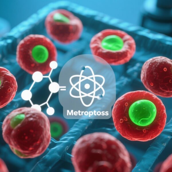
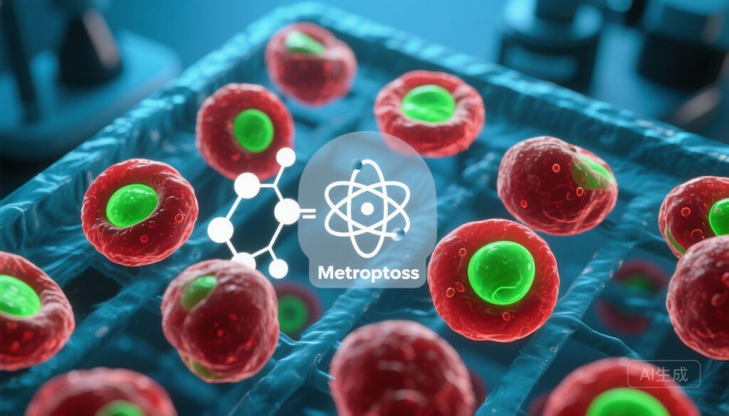
Một minh họa khoa học chi tiết cao: các tế bào bệnh bạch cầu cấp tính myeloid được nuôi cấy dưới kính hiển vi huỳnh quang hiển thị các hạt lipid sáng (màu đỏ) và các dấu hiệu ROS/peroxidation lipid (màu xanh lá cây), phân tử metformin được vẽ mờ ở góc, biểu tượng nguyên tử sắt và các nhãn DGAT1/CD36 nhỏ; nền phòng thí nghiệm mát mẻ, độ tương phản cao, phong cách biên tập hiện đại phù hợp cho bìa tạp chí y học.

Metformin Triggers Ferroptosis in AML Through Lipid Remodeling: A Repurposing Opportunity for Metabolic Subtypes

Metformin Triggers Ferroptosis in AML Through Lipid Remodeling: A Repurposing Opportunity for Metabolic Subtypes

Highlight

– Metformin induces reactive oxygen species (ROS) and ferroptotic cell death in primary AML samples ex vivo, with greatest effect in cases having disrupted lipid metabolism (notably IDH2- and FLT3-mutant disease).

– Lipidomic remodeling with increased triglycerides and polyunsaturated fatty acids (PUFAs), upregulation of lipid-droplet genes including DGAT1, and CD36-mediated uptake are key determinants of metformin sensitivity.

– DGAT1 inhibition synergizes with metformin; iron chelators antagonize its effect—supporting ferroptosis as the operative cell-death mechanism and pointing to rational combination strategies.

Background

Metabolic reprogramming is a hallmark of cancer and a recognized therapeutic vulnerability in acute myeloid leukemia (AML). Many AML cells and leukemic stem cells rely on oxidative phosphorylation (OXPHOS) to sustain survival and chemoresistance, and targeting mitochondrial metabolism has emerged as a promising strategy in preclinical studies.[3] However, many potent OXPHOS inhibitors have dose-limiting toxicities that hinder clinical translation. Metformin, a widely used and generally safe antidiabetic drug, inhibits mitochondrial complex I and reduces OXPHOS at pharmacologic and higher concentrations. Interest in repurposing metformin for oncology rests on its safety profile and pleiotropic metabolic effects, but mechanisms of anti-leukemic activity and predictive biomarkers for response are incompletely defined.

Study design

The study by Sternadt and colleagues (Blood Advances, 2025)[1] interrogated metformin sensitivity across a genetically diverse panel of primary AML samples. Investigators isolated CD34+/CD117+ blasts from clinical specimens and applied label‑free quantitative proteomics. They performed single-sample gene set enrichment analysis (ssGSEA) focusing on metabolic pathways and correlated enrichment scores with ex vivo metformin sensitivity. Functional assays measured reactive oxygen species (ROS), cell death modality, and the effects of lipid manipulations. Lipidomic profiling assessed fatty acid and triglyceride changes after treatment. Genetic perturbations (CD36 knockdown) and pharmacologic modulators (palmitate, DGAT1 inhibitor, iron chelators) were used to dissect mechanism and synergy.

Key findings

The report combines proteomics, functional assays, and lipidomics to generate a coherent mechanistic story. The principal findings are:

1. Metformin triggers ROS generation and ferroptosis in primary AML ex vivo

Ex vivo metformin treatment increased cellular ROS and induced a form of non-apoptotic, iron-dependent cell death consistent with ferroptosis. Ferroptosis is characterized by iron-dependent lipid peroxidation and is distinct from apoptosis and necroptosis.[2] The cell death was mitigated by iron chelation, corroborating iron dependence, and by interventions that limit lipid peroxidation—supporting ferroptosis as the dominant mechanism.

2. Sensitivity correlates with disturbed lipid metabolism and specific genotypes

Proteomic signatures and ssGSEA showed that AML samples with enriched lipid metabolic pathways were more susceptible to metformin. IDH2- and FLT3-mutant samples were particularly sensitive. IDH mutations are known to alter cellular metabolism and redox balance through production of the oncometabolite 2-hydroxyglutarate, which can influence NADPH and lipid handling; FLT3 mutations are also linked to metabolic reprogramming—providing biological plausibility for genotype-associated vulnerability.[4]

3. Lipid uptake and remodeling modify metformin response

Supplementation with palmitate (a saturated fatty acid) enhanced metformin sensitivity in IDH2-mutant cells, while CD36 knockdown reduced sensitivity, indicating that fatty acid uptake via CD36 contributes to metformin-induced ferroptosis. Lipidomic analysis revealed increases in triglycerides and polyunsaturated fatty acids (PUFAs) after metformin exposure, suggesting active lipid remodeling that increases pools susceptible to peroxidation.

4. Lipid droplet machinery is engaged and DGAT1 is a potential combinatorial target

Metformin upregulated genes involved in lipid droplet formation, including DGAT1—an enzyme catalyzing final triglyceride synthesis and lipid droplet biogenesis. Pharmacologic inhibition of DGAT1 produced strong synergy with metformin, amplifying cell death, consistent with the concept that blocking protective lipid storage or altering lipid partitioning can sensitize cells to ferroptosis.

5. Iron chelation antagonizes metformin’s effect

Iron chelators reduced metformin-induced cell death, reinforcing the requirement for iron in the observed ferroptotic mechanism and highlighting potential therapeutic interactions—e.g., patients on iron chelation might derive less benefit if ferroptosis induction is a desired mechanism.

Mechanistic interpretation

Metformin’s primary mitochondrial effect—complex I inhibition—reduces NAD+/NADH turnover and impairs electron transport, leading to increased mitochondrial ROS in susceptible cells. Excess ROS can initiate lipid peroxidation when cellular pools of PUFAs and accessible iron are present, culminating in ferroptotic death.[2] The study indicates that AML cells with active lipid uptake and remodeling pathways accumulate PUFA-containing lipids and triglycerides, creating a substrate pool for peroxidation. DGAT1-mediated triglyceride formation and lipid droplet dynamics may represent protective or buffering mechanisms; inhibiting DGAT1 prevents sequestration of harmful lipid species, thus amplifying ferroptosis. CD36-mediated uptake of exogenous fatty acids, especially saturated FAs such as palmitate, modulates membrane composition and could influence the ratio of saturated to unsaturated lipids, thereby affecting susceptibility to peroxidation. This integrated lipid-centric view links mitochondrial inhibition to ferroptotic death via substrate availability and storage dynamics.

Clinical and translational implications

The work suggests several clinically relevant points:

  • Biomarker-driven repurposing: Proteomic or transcriptomic signatures of lipid metabolism, or specific genotypes (IDH2, FLT3), may identify AML patients more likely to respond to metformin-based strategies.
  • Combination strategies: Co-targeting lipid handling (DGAT1 inhibitors), fatty acid uptake (CD36 antagonists), or exploiting iron metabolism could potentiate metformin efficacy. Conversely, iron chelation could blunt benefit and should be considered when designing trials.
  • Dose and pharmacokinetics caution: Many in vitro studies use metformin concentrations higher than achievable plasma levels with standard dosing. Translational development will require careful dose-finding, potentially localized delivery, or combinations that lower the required metformin exposure.
  • Patient selection and safety: Given metformin’s safety record in diabetes, rapid translation is attractive, but AML patients have comorbidities (renal dysfunction, lactic acidosis risk) that must be accounted for—especially if higher-than-usual doses or combinations are used.

Expert commentary and limitations

This study elegantly connects mitochondrial inhibition to ferroptosis through lipidomic remodeling, strengthening the conceptual framework for targeting metabolic vulnerabilities in AML. Prior work has established OXPHOS dependence in certain AML subsets and highlighted the promise of mitochondrial targeting.[3] The demonstration that metformin—an approved, well-tolerated agent—can engage ferroptosis in primary AML is compelling, particularly because it identifies tractable biomarkers (lipid signatures, DGAT1, CD36) and actionable combinations.

However, limitations must be stressed. The bulk of the data are ex vivo and rely on cellular and biochemical assays; in vivo validation in animal models and patient-derived xenografts is necessary to assess pharmacokinetics, tumor microenvironment effects, and tolerability at effective doses. Metformin’s in vitro active concentrations often exceed therapeutic plasma levels, so combinatorial partners that lower the effective dose will be critical for clinical translation. Heterogeneity of patient samples and complexity of lipid metabolism across patients may complicate biomarker development. Finally, ferroptosis is a context-dependent process and interactions with host iron metabolism, inflammation, and concurrent medications (e.g., iron chelators) require systematic evaluation.

Next steps and research priorities

Priority actions to translate these findings include:

  • Preclinical in vivo studies testing metformin alone and in rational combinations (DGAT1 inhibitors, CD36 modulators) using orthotopic AML models and patient-derived xenografts, with attention to dose, exposure, and toxicity.
  • Development and validation of predictive biomarkers (proteomic lipid signatures, DGAT1/CD36 expression, and mutational status like IDH2/FLT3) that can stratify patients for early-phase trials.
  • Phase I/II biomarker-driven clinical trials assessing safety, pharmacodynamics (ROS, lipid peroxidation markers), and early efficacy signals, while monitoring interactions with iron metabolism and standard AML therapies.
  • Mechanistic studies to map how lipid droplet dynamics and specific lipid species dictate ferroptosis susceptibility in different AML subclones, including leukemic stem cells.

Conclusion

The study by Sternadt et al. positions metformin as a potential metabolic inducer of ferroptosis in AML, with sensitivity shaped by lipid uptake, remodeling, and storage pathways. The data support a precision-medicine approach—using metabolic and genetic biomarkers to select patients and combining metformin with agents targeting lipid handling to amplify ferroptotic death. While promising, the translational path requires rigorous in vivo validation, careful dosing strategies, and early-phase trials to establish safety and proof-of-concept in patients.

Funding and clinicaltrials.gov

The original Blood Advances manuscript reports the study details and author list (Sternadt et al.); specific funding sources and trial registrations should be referenced in the primary publication. As of this writing, clinical trials repurposing metformin for AML driven by ferroptosis have not been widely reported; prospective clinical translation would benefit from registration and biomarker-guided trial design on ClinicalTrials.gov.

References

[1] Sternadt D, Pereira-Martins DA, Chatzikyriakou P, Albuquerque-Simões L, Yang M, Silveira DR, Wierenga AT, Weinhäuser I, Hogeling SM, Oudejans L, Casares-Alaez P, Sarry JE, Frezza C, Huls GA, Quek L, Schuringa JJ. Metformin induces ferroptosis associated with lipidomic remodeling in AML. Blood Adv. 2025 Oct 31:bloodadvances.2025016155. doi: 10.1182/bloodadvances.2025016155. Epub ahead of print. PMID: 41172231.

[2] Dixon SJ, Lemberg KM, Lamprecht MR, Skouta R, Zaitsev EM, Gleason CE, et al. Ferroptosis: an iron-dependent form of nonapoptotic cell death. Cell. 2012;149(5):1060–72.

[3] Lagadinou ED, Sach A, Callahan K, Rossi RM, Neering SJ, Minhajuddin M, et al. BCL-2 inhibition targets oxidative phosphorylation and eradicates quiescent leukemia stem cells. Cell Stem Cell. 2013;12(3):329–341.

[4] Figueroa ME, Abdel-Wahab O, Lu C, Ward PS, Patel J, Shih A, et al. Leukemic IDH1 and IDH2 mutations result in a hypermethylation phenotype, disrupt TET2 function, and block hematopoietic differentiation. Cancer Cell. 2010;18(6):553–567.

AI-friendly thumbnail prompt

A high-detail scientific illustration: cultured acute myeloid leukemia cells under a fluorescence microscope showing bright lipid droplets (red) and ROS/lipid peroxidation markers (green), a stylized metformin molecule semi-transparently overlaid in the corner, an iron atom icon and small DGAT1/CD36 labels; cool laboratory background, high contrast, modern editorial style suitable for a medical journal cover.

Comments

No comments yet. Why don’t you start the discussion?

Để lại một bình luận