Highlight
- Alopecia areata (AA) patients show nearly double the risk of developing microscopic colitis (MC), including lymphocytic and collagenous subtypes.
- AA is also associated with significantly increased risks of other immune-mediated gastrointestinal (GI) disorders, such as celiac disease, Crohn’s disease, eosinophilic esophagitis, and ulcerative colitis.
- Patients with coexisting AA and MC present at a younger age compared to those with MC alone, suggesting potential early immunological interactions.
- The findings emphasize the importance of routine GI screening and gastroenterology referrals in AA patients presenting with chronic digestive symptoms.
Study Background and Disease Burden
Alopecia areata (AA) is an autoimmune condition characterized by non-scarring hair loss resulting from immune-mediated disruption of hair follicles. Although classically regarded as a dermatologic disease, emerging evidence points to systemic immune dysregulation and associations with other autoimmune diseases. Microscopic colitis (MC) is an inflammatory bowel condition subdivided into lymphocytic and collagenous colitis, manifesting as chronic watery diarrhea and thought to be immune-mediated. Despite distinct clinical presentations, both disorders involve aberrant immune responses.
Existing epidemiological data have suggested overlaps between autoimmune dermatologic conditions and gastrointestinal (GI) immune-mediated diseases, but robust large-scale studies delineating these relationships are limited. Understanding AA’s association with GI disorders carries clinical relevance as it may influence early diagnosis, management, and multidisciplinary care pathways. The study by Hirpara et al. endeavors to clarify the risk relationships between AA and immune-mediated GI conditions using a large global dataset.
Study Design
The investigators performed a retrospective cohort analysis utilizing the TriNetX global research network database. The study population included 117,674 patients diagnosed with AA, with a mean age of 33.5 years and predominantly female composition (60.9%). Each AA patient was matched 1:1 with a control subject by age, sex, and race (48.7% White, 14.5% Black, 7.35% Asian).
The primary outcome assessed was the prevalence of immune-mediated gastrointestinal illnesses among those with AA compared to matched controls. These GI conditions were selected based on their known autoimmune or immune-driven pathology and comprised microscopic colitis (including lymphocytic and collagenous colitis), celiac disease, Crohn’s disease, eosinophilic esophagitis, and ulcerative colitis.
Key Findings
The study found that patients with AA had a significantly higher risk of MC compared to those without AA, with an odds ratio (OR) of 1.88 (P < .001). This elevated risk encompassed both major MC subtypes: lymphocytic colitis (OR 1.83; P < .001) and collagenous colitis (OR 1.80; P = .003). Notably, patients presenting with both AA and MC were younger on average (mean age 59.6 years) than those with MC alone (mean age 62.8 years; P = .003), which might indicate earlier onset or a shared immunopathogenic mechanism.
Beyond MC, the AA cohort also demonstrated significantly increased risks for other GI autoimmune diseases:
- Celiac disease (OR 1.87; P < .001)
- Crohn’s disease (OR 1.75; P < .001)
- Eosinophilic esophagitis (OR 1.59; P < .001)
- Ulcerative colitis (OR 1.38; P < .001)
These data indicate a broader association between AA and a spectrum of immune-mediated gastrointestinal disorders.
Expert Commentary
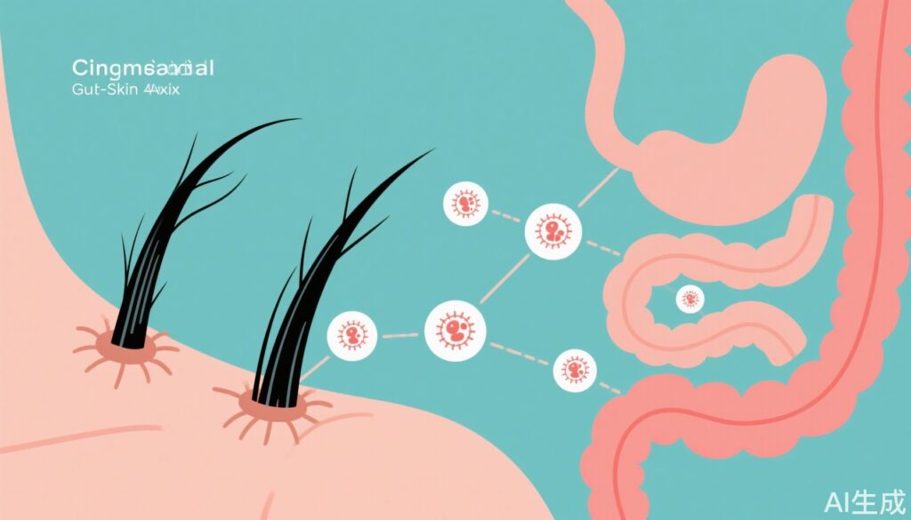
The findings by Hirpara and colleagues contribute to a growing body of evidence supporting a ‘gut-skin axis’ in autoimmune diseases, where immune dysregulation in the gastrointestinal tract may influence distant organ systems such as the skin and hair follicles. The shared increased risk of both lymphocytic and collagenous colitis in AA patients emphasizes potential common pathogenic immune pathways, possibly involving T-cell dysregulation, altered mucosal immunity, or microbiome influences.
However, the retrospective design introduces inherent limitations including potential confounding factors, reliance on diagnostic coding accuracy, and inability to establish causality. Additionally, the TriNetX database does not provide granular clinical detail on disease severity, treatment, or lifestyle factors that could modify risk.
Future prospective studies are warranted to elucidate mechanistic links and temporal relationships, as well as to assess therapeutic implications. From a clinical perspective, this evidence supports the proactive evaluation of GI symptoms in AA patients and integration of dermatologic and gastroenterologic care.
Conclusion
This large-scale retrospective study robustly links alopecia areata with increased risks of microscopic colitis and other immune-mediated gastrointestinal disorders, highlighting a clinically significant gut-skin immunological connection. Clinicians managing AA should maintain a high index of suspicion for concomitant GI autoimmune diseases, particularly in patients presenting with digestive complaints. Screening protocols and timely gastroenterology referrals may facilitate early diagnosis and holistic management.
Further research is needed to clarify underlying immunopathogenic mechanisms and to develop integrated treatment strategies targeting both skin and gut immune dysregulation. This study reinforces the importance of multidisciplinary approaches in autoimmune disease care and augments our understanding of systemic manifestations beyond the primary target organ in AA.