Highlight
1. Tumor-infiltrating lymphocytes (TILs) serve as a critical biomarker influencing melanoma prognosis and immunotherapy response.
2. Traditional TIL assessment by pathologists on hematoxylin and eosin (H&E) stained slides exhibits considerable interobserver variability.
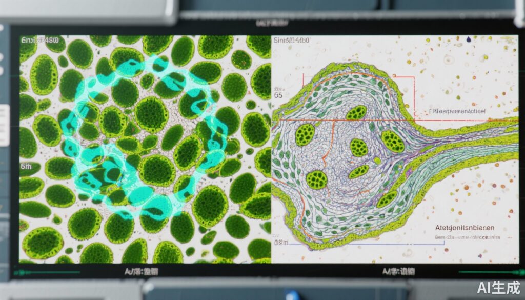
3. An artificial intelligence (AI)-driven TIL quantification algorithm demonstrates superior reproducibility with excellent intraclass correlation coefficients (>0.90).
4. AI-derived TIL scores show significant prognostic associations with survival outcomes, outperforming conventional manual scoring.
Study Background
Melanoma, a highly aggressive skin cancer, presents significant clinical challenges including variable prognosis and heterogeneous response to immunotherapy. Tumor-infiltrating lymphocytes (TILs), immune cells within the tumor microenvironment, have emerged as a provocative biomarker associated with therapeutic outcomes and survival. The degree of TIL infiltration correlates with antitumor immune activity and has prognostic and predictive relevance.
Conventionally, TIL quantification is performed by pathologists assessing hematoxylin and eosin (H&E) stained tissue slides. However, such manual evaluation is subject to interobserver variability, impacted by subjective interpretation of TIL density and distribution, thereby limiting reliability in clinical decision-making. The need for a standardized, reproducible, and scalable TIL assessment method is critical to advance melanoma patient care.
Study Design
This multi-institutional, retrospective prognostic study compared traditional pathologist-read TIL scoring with an AI-driven approach. The study cohort included 111 patients with melanoma, diagnosed between January 2022 and June 2023, from 45 international institutions encompassing academic, clinical, and research settings to capture diverse expertise.
Participants (n=98) in the manual arm were exclusively pathologists, while the AI-assisted arm comprised 11 pathologists and 47 nonpathology scientists. A total of 60 H&E stained melanoma tissue sections underwent TIL evaluation by both manual and AI methods.
Primary outcome measures included reproducibility metrics—intraclass correlation coefficient (ICC) for continuous TIL measures and Kendall’s W for categorical Clark score assessments (brisk, nonbrisk, sparse). Prognostic value of TIL quantification was analyzed using univariable and multivariable Cox regression models adjusted for clinicopathologic variables, applying established dichotomous cutoffs (median and 16.6%) for AI-based TIL percentages.
Key Findings
The AI algorithm exhibited excellent reproducibility with ICC values exceeding 0.90 across all TIL variables, significantly outperforming manual pathologist assessments where ICC reached only 0.61 for stromal TIL counts and Kendall’s W was 0.44 for Clark scoring, indicating moderate reliability.
Prognostically, the AI-derived TIL scores were robustly associated with improved patient outcomes. Using a median cutoff, the hazard ratio (HR) for survival was 0.45 (95% confidence interval [CI], 0.26-0.80; p=0.005), and using the established 16.6% cutoff, the HR was 0.56 (95% CI, 0.32-0.98; p=0.04). This suggests that higher AI-quantified TIL infiltration significantly correlates with better survival.
Conversely, manual TIL measurements demonstrated lower reproducibility and weaker prognostic associations, underscoring challenges of subjective assessment.
The study dataset and AI tool are publicly available, facilitating transparency and further validation by other investigators.
Expert Commentary
This study underscores the transformative potential of artificial intelligence in histopathology, particularly for quantifying complex immune biomarkers such as TILs in melanoma. The high reproducibility and prognostic validity of the AI-driven method address a major limitation of conventional pathology—interobserver variability that can hamper consistent clinical interpretation and management.
Although retrospective design limits direct evidence of clinical utility and impact on treatment decision-making, this work lays a strong foundation for prospective trials evaluating AI-based TIL assessment integration into routine pathology workflows and immunotherapy stratification algorithms.
Future studies should explore AI robustness across diverse patient populations and melanoma subtypes, as well as cost-effectiveness and user training requirements.
Conclusion
The AI algorithm for TIL quantification in melanoma outperforms traditional pathologist assessment in both reproducibility and prognostic significance. This advancement has meaningful implications for improving objective biomarker-based prognostication and guiding immunotherapy decisions.
As the field moves toward precision oncology, AI-driven pathology offers a scalable, standardized approach to harness the prognostic power of the tumor microenvironment. Continued validation and integration into clinical practice will be vital to fully realize this technology’s benefits for melanoma patients.
Funding and Clinical Trials
Funding details were not specified in the provided content. No clinical trial registration information was reported.
References
Aung TN, Liu M, Su D, et al. Pathologist-Read vs AI-Driven Assessment of Tumor-Infiltrating Lymphocytes in Melanoma. JAMA Netw Open. 2025;8(7):e2518906. doi:10.1001/jamanetworkopen.2025.18906. PMID: 40608341; PMCID: PMC12232186.


